Skip Navigation
Skip to contents

Res Vestib Sci : Research in Vestibular Science

OPEN ACCESS
SEARCH
Search

Articles

Page Path
HOME > Res Vestib Sci > Volume 21(1); 2022 > Article
Case Report
전정신경핵 병터에서 발생한 해리성 수직-회선안진
김현성1, 오은혜1, 최재환1,2orcid
Dissociated Vertical-Torsional Nystagmus in Vestibular Nucleus Lesion
Hyun-Sung Kim1, Eun Hye Oh1, Jae-Hwan Choi1,2orcid
Research in Vestibular Science 2022;21(1):19-23.
DOI: https://doi.org/10.21790/rvs.2022.21.1.19
Published online: March 15, 2022

1Department of Neurology, Pusan National University Yangsan Hospital, Yangsan, Korea

2Department of Neurology, Pusan National University College of Medicine, Yangsan, Korea

Corresponding Author: Jae-Hwan Choi Department of Neurology, Pusan National University Yangsan Hospital, 20 Geumo-ro, Mulgeum-eup, Yangsan 50612, Korea Tel: +82-55-360-2122 Fax: +82-55-360-2152 E-mail: rachelbolan@hanmail.net
• Received: February 14, 2022   • Revised: February 23, 2022   • Accepted: March 2, 2022

Copyright © 2022 by The Korean Balance Society. All rights reserved.

This is an open access article distributed under the terms of the Creative Commons Attribution Non-Commercial License (http://creativecommons.org/licenses/by-nc/4.0) which permits unrestricted non-commercial use, distribution, and reproduction in any medium, provided the original work is properly cited.

  • 3,146 Views
  • 77 Download
prev next
해리성 수직-회선안진(dissociated vertical-torsional nystagmus)이란 각 눈에서 관찰되는 수직-회선안진의 크기나 방향이 서로 다른 안진을 일컫는다[1]. 회선성분의 경우 방향은 같고 크기가 서로 다른 반면, 수직성분은 크기와 방향 모두 각 눈에서 다양하게 관찰된다. 즉 양안의 수직 성분 방향이 서로 다른 양상을 보이거나(편측시소안진, hemi-seesaw nystagmus), 모두 상향 또는 하향안진을 보일수 있다. 해리성 수직-회선안진은 안쪽세로다발(medial longitudinal fasciculus)을 침범하는 핵간안근마비(internuclear ophthalmoplegia)나 내측연수 병터에서 주로 보고되고 있으며, 그 병인으로는 안쪽세로다발을 경유하는 수직 전정안구반사나 난형낭안구반사(utriculo-ocular reflex)의 손상이 제시되고 있다[2-4]. 저자들은 안쪽세로다발 침범 없는 전정신경핵(vestibular nucleus) 병터에서 해리성 수직-회선안진을 보인 증례를 경험하였기에 보고하고자 한다.
71세 남자 환자가 내원 당일 갑자기 발생한 회전성 어지럼, 균형 장애, 구음장애로 응급실에 왔다. 어지럼은 체위 변화에 영향을 받지 않았으며 증상은 지속적이었고, 구역, 구토 증상을 동반하였다. 환자는 수십 년 전에 당뇨를 진단받고 약물 복용 중이었다.
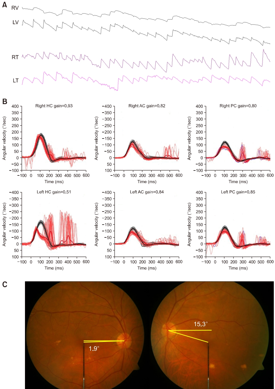
증상 발생 2시간 만에 시행한 신경학적 진찰에서 구음 장애가 관찰되었지만 안구운동장애나 안면근육 마비, 사지 위약감, 감각 저하 등은 없었다. 신경이과적 진찰에서 시계 방향의 회선안진과 상향안진이 관찰되었고, 수평 주시유발안진 역시 관찰되었다. 두부충동검사(head impulse test)에서 좌측으로 두부충동 시 교정성신속안구운동(corrective saccade)이 관찰되었다. 좌측 방향으로 머리가 기울어져 있었고, 교대가림검사(alternate cover test)에서 좌안하사시(hypotropia)가 관찰되었다. 소뇌기능검사에서 사지 실조는 보이지 않았으나 좌측으로 측방돌진(body lateropulsion)이 관찰되었다. 비디오안진검사를 통해 자발안진을 정량적으로 분석한 결과, 양안에서 시계방향으로 향하는 회선안진이 관찰되었지만 우안에서 회선안진이 더 크게 관찰되었고, 상향안진은 주로 좌안에서 관찰되었다 (Fig. 1A). 비디오두부충동검사에서는 좌측 수평반고리관과 양측 뒤반고리관에서 이득(gain)의 저하와 함께 교정성신속안구운동을 보였다(Fig. 1B). 주관적시수직검사에서는 좌측으로 기울임을 보였고(–10.25°; 정상, –3.0° to 3.0°), 안저검사(fundus photography)에서는 좌안에서 외회선(15.3°; 정상, 0°–12.6°)이 관찰되었다(Fig. 1C).
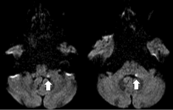
증상 발생 3시간 후 시행한 뇌 자기공명영상의 확산강조영상은 정상이었지만, 2일 후 재시행한 뇌 확산강조영상에서 입쪽 연수(rostral medulla)와 꼬리쪽 교뇌(caudal pons)의 등가쪽 부위(dorsolateral portions)에 급성기 뇌경색 병변이 보였다(Fig. 2). 환자에게 항혈소판제를 투여하였고, 증상 발생 6일 만에 어지럼과 균형장애는 호전되었다.
해리성 수직-회선안진은 주로 핵간안근마비나 내측연수 병터에서 안쪽세로다발의 손상으로 발생한다고 알려져 있다. 그러나 본 증례에서는 해리성 수직-회선안진이 관찰되었지만 안쪽세로다발 손상을 시사하는 안구운동 장애를 보이지 않았으며, 병터 역시 연수-교뇌 이음부의 등가쪽 부위에 위치하고 있어 기존의 보고들과는 차이가 있다. 뇌 자기공명영상에서 관찰되는 병터로 미루어 볼 때 아래소뇌다리(inferior cerebellar peduncle)와 전정신경핵의 손상이 의심되지만, 영상소견만으로는 두 구조물을 구별하기 어렵다. 아래소뇌다리 병터에서는 병측으로 향하는 수평-회선안진과 함께 안구기울임반응이 병터 반대측으로 관찰되며, 두부충동검사는 정상이다[5]. 전정신경핵은 말초 전정신경뿐만 아니라 소뇌와 시각 및 체성감각계로부터 들어온 정보를 처리하고 통합하는 구조물이기 때문에, 전정신경핵 병터에서는 말초 및 중추 전정기능 장애가 함께 나타날 수 있다. 즉 병터 반대측으로 향하는 수평-회선 방향의 자발안진, 병측 두부충동검사 양성 또는 반고리관마비, 병측으로 향하는 안구기울임반응과 같은 말초 전정기능장애와 함께 수평 주시유발안진과 같은 중추 전정기능장애를 보일 수 있다[6]. 더불어 비디오두부충동검사에서는 병측 뿐만 아니라 병터 반대측에서도 전정안구반사의 이득 감소를 보인다. 본 증례에서는 수평 주시유발안진과 함께 안구기울임반응이 병측으로 관찰되고, 비디오두부충동검사에서 병측 수평반고리관과 양측 뒤반고리관의 전정안구반사 장애를 보이고 있어, 아래 소뇌다리보다는 전정신경핵이 주로 침범되었을 것으로 여겨진다.
전정신경핵 병터에서는 대부분 병터 반대측으로 향하는 수평-회선안진과 주시유발안진, 병측 또는 양측으로의 전정안구반사 장애를 보인다[6]. 하지만 자발안진이 병측으로 향하면서 정상 두부충동검사 및 온도안진검사를 보인 증례도 보고되고 있다[7]. 이는 전정신경핵이 꼬리쪽 교뇌와 입쪽 연수에 걸쳐 있는 복합체이며, 상, 하, 외, 내측전정신경핵으로 구분되어 있어, 전정신경핵의 손상 부위나 정도에 따라 신경이과적 소견이 다르게 관찰될 수 있기 때문이다. 대개는 전정신경핵 복합체 중 크기가 가장 큰 내측전정신경핵이 잘 침범되기 때문에 지금까지 주로 보고하고 있는 신경이과적 소견은 내측전정신경핵의 기능 이상으로 인한 안구운동 이상이다[6]. 하지만 내측 전정신경핵 병변이라 하더라도 주시유발안진을 동반하지 않는 경우에는 내측전정신경핵 내 신경적분체(neural integrator) 기능이 정상임을 시사한다[8]. 또한 자발안진이 병측으로 관찰되는 증례에서는 병터가 교뇌 상방에 위치하고 있어 상측전정신경핵의 손상으로 설명하고 있다[7]. 최근에는 비주기교대안진(aperiodic alternating nystagmus) 을 보인 전정신경핵 병터 증례도 보고되었고[9], 본 증례에서도 자발안진이 수평-회선안진이 아닌 해리성 수직-회선안진을 보였다. 이러한 소견들은 전정신경핵 병터에서 병터 반대측으로 향하는 수평-회선안진 외 다양한 양상의 안진을 보일 수 있음을 시사한다.
해리성 수직-회선안진이 발생하는 기전에 대해서는 아직 잘 알려져 있지 않다. 핵간안근마비 33예의 해리성 수직-회선안진을 분석한 연구에 따르면, 병측으로 향하는 회선안진과 비대칭 상향안진(ipsiversive torsional nystagmus with asymmetrical upbeat components)을 보인 경우가 가장 흔하였고, 다음으로 편측시소안진(ipsiversive torsional nystagmus with vertical components in the opposite direction; hemi-seesaw nystagmus), 병측으로 향하는 회선안진과 비대칭 하향안진(ipsiversive torsional nystagmus with asymmetrical downbeat components) 순이었다[2]. 기전으로는 병터 반대측에서 올라오는 수직 전정안구반사의 선택적 손상이나 난형낭안구반사의 이상으로 인한 안구기울임반응, 수직 주시고정(vertical gaze holding)에 관여하는 정중곁로 (paramedian tracts)의 손상 등을 제시하고 있다[1,2]. 본 증례의 경우 핵간안근마비와는 다르게 병터 반대측(시계 방향)으로 향하는 회선안진과 비대칭 상향안진을 보였기 때문에, 병측(좌측) 전정신경핵 내 앞반고리관에서 기원하는 전정안구반사의 선택적 손상을 우선 고려해 볼 수 있다. 이로 인해 앞반고리관에서 병측 눈(좌안)의 상직근과 병터 반대측 눈(우안)의 하사근으로 전달되는 흥분성 구심신호가 전달되지 않아 병측 눈(좌안)에서는 상향안진이, 병터 반대측 눈(우안)에서는 회선안진이 더 크게 관찰될 수 있다. 하지만 비디오두부충동검사에서 양측 앞반고리관의 전정안구반사는 정상이었기 때문에 위 가설로는 안진 양상을 모두 설명할 수 없다.
해리성 수직-회선안진의 기전을 설명할 수 있는 또 다른 가설로는 난형낭안구반사의 이상이다[1,2]. 본 증례에서 병측으로 스큐편위가 관찰되고 주관적시수직이 병측으로 기울어져 있어 난형낭 기능 이상이 있음을 시사한다. 난형낭안구경로(utriculo-ocular pathway)는 난형낭에서 시작하여 동측 전정신경핵, 반대측 안쪽세로다발과 안구 운동신경핵을 거쳐 반대측 Cajal간질핵(interstitial nucleus of Cajal)에 다다르며, 수평 선형가속운동뿐 아니라 중력 수용경로(graviceptive pathway)의 역할을 담당하여 갸웃면 (roll plane)에서 안구 위치를 조절한다[1]. 따라서 전정신경핵 병터로 인한 난형낭안구반사의 이상으로 안구기울임반응이 병측(좌측)으로 발생하고, 이를 교정하기 위한 안구운동인 수직-회선안진이 병터 반대측으로 나타날 수 있다. 과거 동물실험에서 Cajal간질핵에 muscimol을 주입하여 비활성시켰을 때 안구의 회선운동과 함께 수직-회선 안진이 발생함을 증명하였다[10]. 하지만 본 증례의 경우, 안구회선이 주로 관찰되는 좌안에서 오히려 회선안진이 더 작게 관찰되는 점, 우안에서는 하향안진이 관찰되지 않는 점, 그리고 안구기울임반응이 있는 모든 환자에서 해리성 수직-회선안진이 관찰되지 않는 점 등은 난형낭안구반사의 이상으로 모두 설명할 수 없다. 따라서 해리성 수직-회선안진의 기전 규명을 위한 추가적인 연구가 필요하다.

저자들은 이 논문과 관련하여 이해관계의 충돌이 없음을 명시합니다.

본 연구는 2021년도 양산부산대학교병원 임상연구비지원으로 이루어졌음.
Fig. 1.
(A) Eye-movement recordings show asymmetric contraversive (clockwise from the patient’s perspective, right eye>left eye) torsional nystagmus with markedly asymmetric upbeat (left eye>right eye) components. (B) Video head impulse tests show decreased vestibulo-ocular reflex gains for the left horizontal semicircular canal (HC) and both posterior semicircular canals (PCs) with corrective catch-up saccades. (C) Fundus photography demonstrates an extorsion of the left eye (15.3°; normal range, 0°–12.6°). RV, vertical position of the right eye; LV, vertical position of the left eye; RT, torsional position of the right eye; LT, torsional position of the left eye.
rvs-21-1-19f1.jpg
Fig. 2.
Follow-up diffusion-weighted magnetic resonance imaging 2 days later disclose a small acute infarction (arrows) along the dorsolateral portion of the rostral medulla and caudal pons.
rvs-21-1-19f2.jpg
  • 1. Leigh RJ, Zee DS. The neurology of eye movements. 5th ed. Oxford: Oxford University Press; 2015.
  • 2. Jeong SH, Kim EK, Lee J, Choi KD, Kim JS. Patterns of dissociate torsional-vertical nystagmus in internuclear ophthalmoplegia. Ann N Y Acad Sci 2011;1233:271–8.ArticlePubMed
  • 3. Lee SU, Park SH, Jeong SH, Kim HJ, Kim JS. Evolution of torsional-upbeat into hemi-seesaw nystagmus in medial medullary infarction. Clin Neurol Neurosurg 2014;118:80–2.ArticlePubMed
  • 4. Choi KD, Jung D S, Park KP, Jo JW, Kim JS. Bowtie and upbeat nystagmus evolving into hemi-seesaw nystagmus in medial medullary infarction: possible anatomic mechanisms. Neurology 2004;62:663–5.ArticlePubMed
  • 5. Choi JH, Seo JD, Choi Y R, Kim MJ, Kim HJ, Kim JS, et al. Inferior cerebellar peduncular lesion causes a distinct vestibular syndrome. Eur J Neurol 2015;22:1062–7.ArticlePubMed
  • 6. Kim HJ, Lee SH, Park JH, Choi JY, Kim JS. Isolated vestibular nuclear infarction: report of two cases and review of the literature. J Neurol 2014;261:121–9.ArticlePubMed
  • 7. Chang TP, Wu YC. A tiny infarct on the dorsolateral pons mimicking vestibular neuritis. Laryngoscope 2010;120:2336–8.ArticlePubMed
  • 8. Ataç C, Kısabay A, Çetin Akkoç C, Saruhan G, Çelebisoy N. Vestibular nuclear infarction: case series and review of the literature. J Stroke Cerebrovasc Dis 2020;29:104937. ArticlePubMed
  • 9. Kim CH, Choi KD. Aperiodic alternating nystagmus in isolated vestibular nucleus infarction. J Neurol 2019;266:2875–7.ArticlePubMed
  • 10. D as V E, Leigh RJ, Sw ann M, Thurtell MJ. Muscimol inactivation caudal to the interstitial nucleus of Cajal induces hemi-seesaw nystagmus. Exp Brain Res 2010;205:405–13.ArticlePubMedPMC

Figure & Data

References

    Citations

    Citations to this article as recorded by  

      • PubReader PubReader
      • ePub LinkePub Link
      • Cite
        CITE
        export Copy
        Close
        Download Citation
        Download a citation file in RIS format that can be imported by all major citation management software, including EndNote, ProCite, RefWorks, and Reference Manager.

        Format:
        • RIS — For EndNote, ProCite, RefWorks, and most other reference management software
        • BibTeX — For JabRef, BibDesk, and other BibTeX-specific software
        Include:
        • Citation for the content below
        Dissociated Vertical-Torsional Nystagmus in Vestibular Nucleus Lesion
        Res Vestib Sci. 2022;21(1):19-23.   Published online March 15, 2022
        Close
      • XML DownloadXML Download
      Figure
      • 0
      • 1
      Related articles
      Dissociated Vertical-Torsional Nystagmus in Vestibular Nucleus Lesion
      Image Image
      Fig. 1. (A) Eye-movement recordings show asymmetric contraversive (clockwise from the patient’s perspective, right eye>left eye) torsional nystagmus with markedly asymmetric upbeat (left eye>right eye) components. (B) Video head impulse tests show decreased vestibulo-ocular reflex gains for the left horizontal semicircular canal (HC) and both posterior semicircular canals (PCs) with corrective catch-up saccades. (C) Fundus photography demonstrates an extorsion of the left eye (15.3°; normal range, 0°–12.6°). RV, vertical position of the right eye; LV, vertical position of the left eye; RT, torsional position of the right eye; LT, torsional position of the left eye.
      Fig. 2. Follow-up diffusion-weighted magnetic resonance imaging 2 days later disclose a small acute infarction (arrows) along the dorsolateral portion of the rostral medulla and caudal pons.
      Dissociated Vertical-Torsional Nystagmus in Vestibular Nucleus Lesion

      Res Vestib Sci : Research in Vestibular Science
      TOP