Skip Navigation
Skip to contents

Res Vestib Sci : Research in Vestibular Science

OPEN ACCESS
SEARCH
Search

Articles

Page Path
HOME > Res Vestib Sci > Volume 21(4); 2022 > Article
Video
고개 회전 시 특징적인 안진을 보이는 척추동맥압박증후군 1예
전은주orcid
A Case of Vertebral Artery Compression Syndrome Showing Characteristic Nystagmus during Head Rotation
Eun-Ju Jeonorcid
Research in Vestibular Science 2022;21(4):111-112.
DOI: https://doi.org/10.21790/rvs.2022.21.4.111
Published online: December 15, 2022

Department of Otolaryngology, Incheon St. Mary’s Hospital, College of Medicine, The Catholic University of Korea, Incheon, Korea

Corresponding Author: Eun-Ju Jeon Department of Otolaryngology, Incheon St. Mary’s Hospital, College of Medicine, The Catholic University of Korea, 56 Dongsu-ro, Bupyeong-gu, Incheon 21431, Korea Tel: +82-32-280-7372, Fax: +82-50-4411-7964, E-mail: ejmercy@catholic.ac.kr
• Received: November 9, 2022   • Revised: November 8, 2022   • Accepted: December 2, 2022

Copyright © 2022 by The Korean Balance Society.

This is an open access article distributed under the terms of the Creative Commons Attribution Non-Commercial License (http://creativecommons.org/licenses/by-nc/4.0) which permits unrestricted non-commercial use, distribution, and reproduction in any medium, provided the original work is properly cited.

  • 2,345 Views
  • 91 Download
A 71-year-old man visited the clinic with a complaint of vertigo that had persisted since the past 2 months. The patient reported that upon turning his head to the left or tilting it backwards, he experienced vertigo, which persisted and worsened until the head was brought back to the straight position. He further complained of tinnitus on the right side that persisted since the development of dizziness. He had undergone brain magnetic resonance imaging (MRI)+magnetic resonance angiography+diffusion to evaluate the tinnitus in his right ear at the neurosurgery department 3 years ago. In these scans, hypoplasia of the right vertebral artery was observed. The examination of nystagmus using Video-Frenzel glasses (SLMED Co., Seoul, Korea) revealed that when the head was turned to the right in the sitting position, there was no dizziness and nystagmus, and upon turning the head to the left, rotational vertigo and left torsional and downward nystagmus were observed. In the Dix-Hallpike position test, there was no nystagmus on the right side, whereas the same nystagmus and dizziness were induced on the left side upon turning the head to the left in the sitting position (Supplementary Video 1 ). He further complained that tinnitus on the right side aggravated when the head was turned to the left, which caused vertigo and nystagmus. In the supine roll test, nystagmus was not induced on either sides.
Cervical spine MRI revealed multiple bony spurs, diffuse posterior bulging discs, and degenerative changes in the C3–T1 spinal segments (Fig. 1). Vertebral artery compression syndrome was confirmed by the observation of compression of the left vertebral artery by the bony spur of the cervical vertebra (C5–6 level) and the rapid decrease of the cerebral blood flow when the head was turned to the left on dynamic four-vessel angiography (Fig. 2). When the head was turned to the left, the basilar artery flow decreased but was maintained. Based on the nystagmus pattern (mixed downbeat, torsional, and horizontal) and accompanying tinnitus in the rotated head position, the vertigo and nystagmus in vertebral artery compression syndrome may be resulted from transient ischemia in the peripheral labyrinth. However, the prominent downbeat nystagmus can be also explained by transient ischemia of the inferior cerebellum. Written informed consent was obtained for publication of this case report and accompanying images.
This report was approved by the Institutional Review Board of Incheon St. Mary’s Hospital (No. OC22ZASI0141).
Supplementary materials can be found via https://doi.org/10. 21790/rvs.2022.21.4.111.
Supplementary Video 1.
Video-Frenzel glass examination revealed left torsional and downbeat nystagmus that was induced upon head rotation in the sitting position, which is characteristic of the vertebral artery compression syndrome.

CONFLICT OF INTEREST

No potential conflict of interest relevant to this article was reported.

FUNDING/SUPPORT

None.

Fig. 1.
Cervical spine magnetic resonance imaging revealed the protrusion of a bony spur (arrow) that compressed the course of the left vertebral artery.
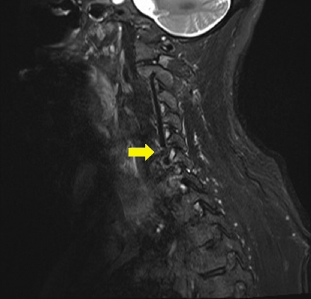
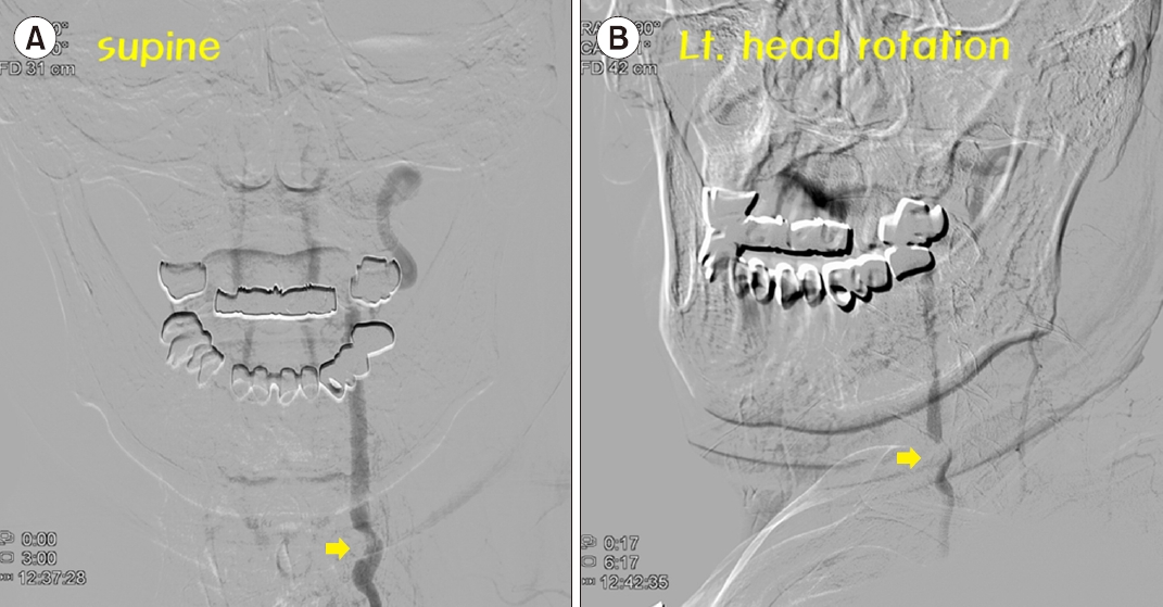
rvs-21-4-111f1.jpg
Fig. 2.
Dynamic four-vessel angiography. (A) Compression of the left vertebral artery by the bony spur (arrow) of the cervical vertebra and (B) rapid decrease in the cerebral blood flow when the head was turned to the left (arrow).
rvs-21-4-111f2.jpg
  • 1. Kim JS, Newman-Toker DE, Kerber KA, Jahn K, Bertholon P, Waterston J, et al. Vascular vertigo and dizziness: diagnostic criteria. J Vestib Res 2022;32:205–22.ArticlePubMedPMC
  • 2. Choi KD, Choi JH, Kim JS, Kim HJ, Kim MJ, Lee TH, et al. Rotational vertebral artery occlusion: mechanisms and long-term outcome. Stroke 2013;44:1817–24.ArticlePubMed

Figure & Data

References

    Citations

    Citations to this article as recorded by  

      • PubReader PubReader
      • ePub LinkePub Link
      • Cite
        CITE
        export Copy
        Close
        Download Citation
        Download a citation file in RIS format that can be imported by all major citation management software, including EndNote, ProCite, RefWorks, and Reference Manager.

        Format:
        • RIS — For EndNote, ProCite, RefWorks, and most other reference management software
        • BibTeX — For JabRef, BibDesk, and other BibTeX-specific software
        Include:
        • Citation for the content below
        A Case of Vertebral Artery Compression Syndrome Showing Characteristic Nystagmus during Head Rotation
        Res Vestib Sci. 2022;21(4):111-112.   Published online December 15, 2022
        Close
      • XML DownloadXML Download
      Figure
      We recommend
      Related articles

      Res Vestib Sci : Research in Vestibular Science