Skip Navigation
Skip to contents

Res Vestib Sci : Research in Vestibular Science

OPEN ACCESS
SEARCH
Search

Articles

Page Path
HOME > Res Vestib Sci > Volume 16(1); 2017 > Article
Review
메니에르병 병인기전의 전기생리학적 연구
김성헌
Ménièreʼs Disease and Electrophysiology
Sung Huhn Kim
Research in Vestibular Science 2017;16(1):1-9.
DOI: https://doi.org/10.21790/rvs.2017.16.1.1
Published online: March 15, 2017

Department of Otorhinolaryngology, Yonsei University College of Medicine, Seoul, Korea

Corresponding Author: Sung Huhn Kim Department of Otorhinolaryngology, Yonsei University College of Medicine, 50 Yonsei-ro, Seodaemun-gu, Seoul 03722, Korea Tel: +82-2-2228-3604 Fax: +82-2-393-0580 E-mail: fledermaus@yuhs.ac
• Received: February 20, 2017   • Revised: February 22, 2017   • Accepted: February 22, 2017

Copyright © 2017 by The Korean Balance Society. All rights reserved.

This is an open access article distributed under the terms of the Creative Commons Attribution Non-Commercial License (http://creativecommons.org/licenses/by-nc/4.0) which permits unrestricted non-commercial use, distribution, and reproduction in any medium, provided the original work is properly cited.

  • 11,699 Views
  • 178 Download
next
  • Inner ear is composed of cochlea, vestibule, and endolymphatic sac which are enclosed by thin layer of epithelial cells. The enclosed space is filled with fluid named as endolymph where the [K] is high and [Na] is low. This unique ion composition is very important in maintaining normal hearing and balance function by providing K ions into sensory hair cells, which finally depolarize hair cells to facilitate the transport of sound and acceleration stimulation to central nervous system. The ion composition of inner ear is maintained by various ion transport through ion channels, transporters, and exchangers in the inner ear sensory and extra-sensory epithelium. The disruption of normal endolymphatic ion composition by the deterioration of the function of those ion channels can cause dysfunction of sensory epithelium, which consequently results in hearing and balance disorders. One of the possible pathology from the disruption of inner ear ion homeostasis is endolymphatic hydrops which is a phenomenon of excessive fluid accumulation of inner ear. The dysfunction of ion channels in inner ear epithelium can be an etiology of Ménièreʼs disease since endolymphatic hydrops is a main pathological finding of the disease. In this review, we discussed about the possible pathological mechanism of Ménièreʼs disease as a perspective of channelopathy as well as the role of various ion channels in the regulation of inner ear fluid volume based on the findings revealed by electrophysiological studies.
메니에르병은 내이의 내림프액이 과다하게 축적되어 내림프 공간이 팽창되는 현상인 내림프수종(endolymphatic hydrops)이 원인 병리현상으로 알려져 있다[1]. 내림프수종을 유발하는 원인은 명확하지 않지만, 내림프 부피를 조절하는 기전의 이상으로 인한 내림프의 과다 생성이나 흡수부전에 의해 발생된다고 생각된다[2]. 메니에르병에 대한 기초연구는 대부분 내림프수종의 발생, 진단 및 치료 등 광범위한 분야에서 시행되었으나, 어떠한 연구도 명확한 해답을 찾지 못했다. 내림프수종의 발생기전을 규명하기 위해서는 이상 단백이나 유전자의 발현 등을 연구하는 일반적인 분자생물학적 기법 외에 실제로 병인기전과 연관된 현상을 관찰하는 기능적인 연구가 동반되는 것이 중요하다. 하지만, 아직 메니에르병의 명확한 동물모델이 없고, 메니에르병 환자에서 실험을 위한 내이 시료를 취득하는 것에 한계가 있어 기능적인 실험이 잘 시행되지 못하였다.
내이의 기초 생리 및 내림프수종의 병인기전 연구에 활용되는 대표적인 기능적 연구방법이 전기생리학이다. 내이의 대부분은 내림프액과 외림프액이라는 액체로 구성되어 있고, 여기에는 많은 이온들이 포함되어 있어, 특정한 이온의 이동과 이로 인해 유발되는 이온농도의 변화가 내림프의 부피변화를 초래하기 때문에, 내이의 이온 변화를 연구하는 전기생리학은 내림프수종의 기초연구에 중요한 역할을 한다. 본 종설에서는 메니에르병의 근거가 될 수 있는 이온수송에 관한 기초 전기생리학적연구 결과들을 고찰하여 그 병리기전을 유추하고 향후 연구방향을 제시하고자 한다.
1. 내이의 해부학적 구조 및 내/외림프액의 이온조성
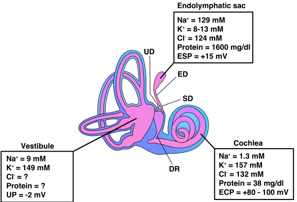
사람 내이는 측두골의 추체골 내부에 위치하며, 청각을 담당하는 달팽이관(cochlea)과 평형기능을 담당하는 전정기관(vestibule), 그리고 전정기관에서 내림프낭관(endolymphatic duct)을 통해 연결된 내림프낭(endolymphatic sacz) 으로 구성된다(Figure 1). 추체골 내부의 이들 구조는 얇은 상피세포막으로 둘러싸여 있으며, 이를 막성미로(membranous labyrinth)라고 하고, 막성미로를 둘러싸는 골부를 골성미로(bony labyrinth)라고 한다. 막성미로의 내부는 높은 K 농도와 낮은 Na 농도의 내림프액(endolymph)이 채우고 있으며, 막성미로와 골성미로 사이에는 이와 반대로 높은 Na 농도와 낮은 K 농도의 외림프액(perilymph)가 채우고 있다(Figure 1) [3]. 하지만, 내림프낭의 내림프는 예외적으로 높은 Na 농도와 낮은 K 농도의 조성을 가지고 있으며, 달팽이관과 전정기관의 내림프에 비해 단백질의 함유량이 높다(Figure 1) [3]. 내림프액 및 외림프액의 특이한 조성의 이온농도는 막성미로의 각종 상피세포에 위치한 이온채널에 의해서 항상 일정하게 조절되며, 이를 내이의 이온항성성(inner ear homeostasis)이라 한다. 청각 및 가속도 자극은 달팽이관과 전정기관 유모세포(hair cell)의 입체섬모(stereocilia)에 위치한 물리적민감성비선택적양이온채널(mechano-sensitive non-selective cation channel)을 통하여 K 이온을 유입시켜 청신경과 전정신경말단부의 탈분극을 유도하고, 이를 통하여 해당 자극이 중추신경계로 전달되는데, 유모세포 입체섬모의 이온채널은 모든 양이온을 비선택적으로 흡수할 수 있어, 내림프의 높은 K 이온농도는 정상적인 유모세포의 기능을 유지하는데 필수적이다[4]. 만약, 내림프의 Na이나 Ca 등의 양이온 농도가 K보다 월등히 높을 경우, 이들 이온이 입체섬모의 이온채널을 통해 더 많이 흡수되어 유모세포 내에 축적되어 정상적인 탈분극을 유도하지 못하고 세포의 손상을 초래한다. 즉, 다른 세포외액과는 다른 내림프액의 특이 이온조성은 청력과 평형기능의 유지를 위해서는 필수적이며, 내이항상성이 파괴되는 경우 유모세포의 기능에 이상이 생겨 각종 청력기능과 평형기능 이상이 발생한다. 내이항상성은 정상적인 유모세포 기능을 유지하기 위해서 필수적이지만, 내이의 정상적인 부피를 유지하기 위해서도 내이의 이온농도는 일정하게 유지되어야 한다. 인체에서 물의 이동은 대개 이온의 이동에 수반되며, 이는 내이에서도 동일하다. 물의 이동과 연관된 주요 이온은 Na과 Cl 가 있으며, 해당 이온들이 이온채널을 통해 이동하면, 물은 세포막에 위치한 아쿠아포린(aquaporin)과 같은 수분이동채널이나 세포사이간극을 통하여 부수적으로 이동하게 된다. 비단 Na이나 Cl 뿐만 아니라 다른 이온들의 이동에 의해서도 내림프와 외림프의 삼투압 차이가 발생할 수 있으며, 이 또한 수분 이동을 유발할 수 있다. 내이의 유모세포 이외에 막성 미로를 구성하는 비감각성 상피세포에도 다양한 종류의 이온채널들이 존재하며, 이를 통한 Na, Cl 등의 이동은 내림프액의 부피조절과 연관이 있을 것이다. 때문에, 내이의 정상적인 이온수송에 이상이 발생한 경우 내림프액과 외림프액 간의 이온농도차에 의한 삼투압 변화가 초래되고, 이는 수분의 이동을 초래하여 내림프수종을 유발할 수 있다. 즉, 내이의 비감감성상피세포의 이온채널 혹은 이에 의해 동반되는 수분이동경로에 이상이 발생한 경우 메니에르병이 발생할 수 있다는 가설이 제시되었고[5], 대부분의 전기생리학적 연구는 이러한 가능성에 초점을 맞추고 있다.
2. 메니에르병과 연관된 전기생리학적적 연구
앞에서 기술한 바와 같이 기능적 연구를 위한 메니에르병 환자의 조직 채취가 어렵고, 메니에르병의 명확한 동물 모델이 없어 환자 조직이나 메니에르병 동물모델을 이용한 직접적인 전기생리학적 연구는 매우 드물다. 때문에 동물 정상 내이조직의 이온채널의 분포 및 그 기능을 규명하고, 이와 내림프수종의 연관성을 가설로 제시하는 연구가 대부분이며, 일부에서는 임상적 치료약제가 내이 이온채널의 기능에 미치는 영향을 분석하여 치료기전을 제시하는 연구가 시행되었다.
전기생리학적 연구를 통해 내림프의 부피조절에 관여할 것이라고 생각되는 이온채널은 내이 비감각상피세포의 전반에 걸쳐 분포하고 있으며, 아직도 그 기능은 일부 만이 밝혀졌다. 내림프에 가장 많이 분포하는 K은 달팽이관의 혈관선조(stria vascularis)의 변연세포(marginal cell)와 전정기관 팽대부와 난형낭의 암흑세포(dark cell) 첨부에 위치한 KCNQ1/KCNE1을 통하여 내림프로 배출되며, 소리와 가속도 자극 시 각각 유모세포의 입체섬모에 위치한 비선택적 양이온채널로 흡수된다[6]. 내이에서 K의 배출 및 흡수, 그리고 재활용은 소리 및 가속도 자극을 인지하는데 중요한 역할을 하며, 그 자세한 기전은 이미 여러 논문에서 기술하고 있어 여기서는 따로 기술하지는 않겠다[6]. K의 이동도 내림프 부피 조절에 관여할 수 있으나 아직까지 직접적인 근거는 없으며, K 이온의 이동은 주로 소리와 가속도 자극을 인지하는데 중요한 역할을 할 것이라 생각된다. 또한, 난청을 일으키는 각종 K 이온채널의 유전자 변이 동물모델에서도 내림프수종이나 메니에르병과 유사한 증상을 보이지 않는다. K 이온채널 외 내림프 부피조절에 주로 관여할 것이라 생각되는 이온은 Na과 Cl 가 있으며, 본 종설에서는 이들의 이동과 내림프부피조절 간의 연관성에 대해 더 자세히 살펴보겠다.

1) 내이의 Na 이동과 내림프 부피 조절 간의 연관성

내림프에서 Na의 흡수에 관여하는 이온채널 중 그 기능이 실험을 통해 확인된 것으로는 아밀로라이드 민감성 Na 통로(amiloride-sensitive ion channel), 상피세포 Na 통로(epithelial Na channel, ENaC), Na/H 교환체(Na/H exchanger, NHE), 신장활성양이온통로(stretch-activated ion channel), 퓨린수용체 중 P2X 계열의 이온통로 및 칼슘민감성 비선택적양이온통로(Ca sensitive non-selective cation channel) 등이 있으며, 그 분포부위는 Table 1Figure 2와 같다[7-17]. 이들 이온채널은 모두 내림프의 Na을 흡수하는 기능을 하며, 이 중 신장활성양이온채널과 P2X 수용체, 칼슘민감성 비선택적양이온통로 등은 Na 뿐만 아니라 다른 양이온도 비선택적으로 흡수한다. 특히, 달팽이관에서는 +80–100 mV로 유지되는 와우내전위(endocochlear potential)가 존재하여, Na의 흡수가 더욱 촉진될 것으로 생각된다[18]. 흡수된 Na은 상피세포 기저부의 Na/K–ATPase를 통하여 배출되며, Na/K–ATPase로 흡수된 K은 기저부의 여러 종류의 K 이온 채널을 통하여 배출된다(Figure 2)[18]. Na/K–ATPase는 혈관조의 기저세포와 암흑세포의 기저부에서 Na을 배출하고 K을 흡수하여 해당세포의 첨부에서 K을 배출하는 원동력으로도 작용을 한다(Figure 2)[18]. Na과 K 및 2Cl 를 같이 흡수하는 Na–K–2Cl 공수송체(Na–K–2Cl cotransporter, NKCC)는 혈관조, 암흑세포, 반고리관상피세포와 내림프낭에서는 그 기능이 발견되어, 해당 세포의 기저부에서 Na의 흡수에 관여할 것으로 생각되며[18], 이는 세포첨부의 Na의 흡수보다는 혈관조와 암흑세포에서는 세포첨부에서 K 배출을 위하여 K을 기저막에서 세포 내로 공급하고 반고리관상피세포와 내림프낭에서는 Cl 이온의 배출을 위하여 기저막에서 Cl 를 세포 내로 공급하는 역할을 할 것이라 생각된다(Figure 2). NKCC와 Na/K–ATPase로 흡수된 K은 대개 세포 기저부의 K 통로를 통하여 다시 배출되어 세포 내, 외부의 항상성을 유지한다(Figure 2)[18]. 달팽이관과 전정기관의 내림프에서 Na이온이 적절히 흡수되어 외림프로 배출되지 않을 경우 내림프에 Na이 축적되고, 이는 내림프 부피를 증가시키거나 유모세포로 Na의 공급을 늘려 유모세포의 손상을 유발할 수 있다. 하지만, P2X 수용체의 경우 내이에서 분비되는 ATP에 의해 자극이 되어 주로 유모세포의 주변의 외구세포나 유모세포 주변의 전이세포에서 양이온 흡수를 촉진하는데, 이들 현상은 주로 소음이나 가속도 자극 시 발생하거나, 내이의 발생 기간동안 관찰이 되어, 내림프의 부피조절 보다는 유모세포의 기능을 조절하거나 내이의 보호기전으로 작용할 것으로 생각된다[15,18]. P2X 수용체는 난형낭의 전이세포에서는 Ca2+을 흡수하여 이석의 생성 및 유지에 중요한 기능을 하는 것으로 보고되었다[17]. 라이스너막과 구형낭 지붕의 비감각성상피, 그리고 반고리관상피세포에서 ENaC을 통한 Na의 흡수는 마우스실험에서 부신피질호르몬(glucocorticoid)에 의해 촉진이 되는 것으로 규명되어[9-11], 메니에르병에서 부신피질호르몬제의 사용 근거를 제공하는 여러 가지 가설 중 중요한 한 가지 가설이다. 내림프낭은 K보다 Na 농도가 높은 내림프액으로 채워져 있어 Na의 이동이 달팽이관이나 전정기관 보다 더욱 활발히 일어날 가능성이 높으며, 이는 내림프의 부피조절에 중요한 역할을 할 것이라 제시되었다[18]. 내림프낭은 내이를 구성하는 기관 중 유일하게 비감각성상피로만 구성되며, 이전부터 내이 항상성과 내림프부피조절에 중요한 역할을 할 것이라 생각되었다[3]. 특히, 메니에르병 환자의 측두골 표본을 이용한 병리연구에서 내림프낭의 일부 섬유화 및 면역학적 이상 소견이 발견되어, 내림프낭의 이상이 메니에르병을 초래할 가능성이 있다고 제시되었다[19]. 내림프낭 상피세포는 크게 미토콘드리아가 다수 분포하는 세포(mitochondria-rich cell, MRC)와 리보솜이 다수 분포하는 세포(ribosome-rich cell, RRC)의 두 가지 형태로 나뉘며, Na의 흡수는 MRC에서 주로 일어난다[18]. 내림프낭에서는 +10–15 mV의 양전위의 내림프전위가 유지되어 양이온의 흡수를 촉진시킬 것으로 생각된다[18]. 내림프낭에 이상이 생길 경우 내림프수종이 유발될 수 있을 가능성이 전기생리학적 실험으로 제시되었는데, 염증성 사이토가인 중 interleukin-1β와 interferon-γ는 배양된 사람 내림프낭 상피세포에서 ENaC과 NHE를 통한 Na의 흡수를 저하시켜 면역반응에 의한 내림프낭의 염증반응이 내림프수종을 초래할 가능성이 있다고 제기되었다[12,13].

2) Cl 이온의 이동과 내림프 부피 조절 간의 연관성

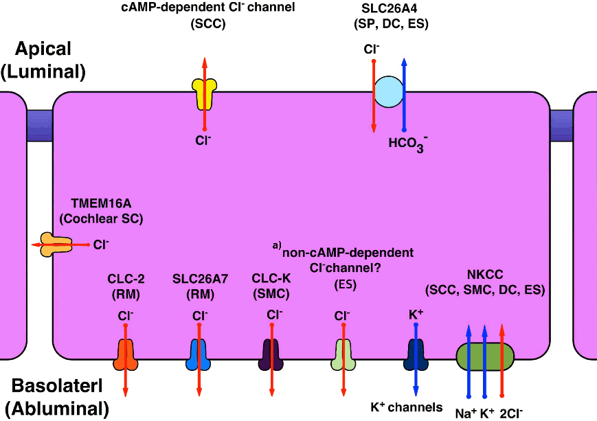
내이에서 Cl 이온에 관여하는 Cl 채널 및 음이온 교환체는 매우 다양하게 확인되었다. 이 중 전기생리학적으로 그 기능이 확인된 것은 CLC-K, CLC-2, SLC26A7, c-AMP의존성 Cl 통로(cAMP-dependent Cl channel), TMEM16A 등의 Cl 채널과 SLC26A4와 같은 음이온 교환체가 있으며, 그 분포 부위는 Table 2Figure 3과 같다[16,20-24]. 달팽이관의 라이스너막에서 CLC-2SCL26A7은 상피세포의 기저부에 분포하여 Cl 이온을 외림프로 배출하여 내림프의 부피조절과 연관성이 있을 것으로 생각되나, 어떠한 경로로 Cl 이온이 세포 내로 들어오는지 밝혀지지 않았다(Figure 3)[21,22]. CLC-K 채널은 혈관선조의 변연세포의 기저부에 분포하며, 여기서 Cl 이온의 이동은 변연세포에서 기저부의 NKCC 와 짝을 이루어 내림프로 K 이온을 배출하는 원동력을 제공하는 역할을 하리라 생각된다(Figure 3)[20]. 또한 TMEM16A는 발생 시에 달팽이관의 지지세포의 기저부에서 Cl 이온을 배출하여 지지세포의 부피를 조절하고, 유모세포의 기능을 조절하는 역할을 할 것으로 생각되나, 내림프부피 조절과의 연관성은 명확하지 않다[16].
전정기관에서는 배양된 쥐의 반고리관 상피세포에서 c-AMP 의존성 Cl 통로에 의해 Cl 이온이 배출됨이 확인되었으며, 이는 제2형 베타교감신경수용체(β2-adrenergic receptor)자극이나 부신피질호르몬제 자극에 의해 촉진된다[23,25]. 상기 결과는 메니에르병이나 돌발성 난청에서 치료제로 사용되는 부신피질호르몬제의 역할에 대한 약리학적 근거가 될 수 있으며, 메니에르병에서 정신적 혹은 육체적인 스트레스로 유발된 교감신경의 활성화 또한 내림프수종에 영향을 미칠 수 있음을 뒷받침한다.
SLC26A4는 HCO3, I, 포르메이트(formate) 등의 음이온을 Cl 와 교환하여 수송하는 음이온교환체(anion exchanger)로 내림프의 pH 조절에 관여하며, 해당 단백은 펜드린(pendrin)이라고 한다[24]. 펜드린은 달팽이관, 전정기관, 내림프낭의 특정부위에 분포하며(Table 2, Figure 3), 펜드린의 기능 이상이 있을 경우 내림프수종에 의한 전정도수관 확장증이 발생하고 난청, 평형기능장애 등이 초래된다. 펜드린 유전자 이상에 의한 선천성 난청은 상염색체열성으로 유전되고, 우리나라에서 가장 흔한 선천성 난청의 원인이다[24,26]. 펜드린 유전자결핍 마우스모델에서 펜드린에 의한 세포 내 HCO3 와 내림프의 Cl 교환이 이루어지지 않아 내림프가 산성화 되고, 이는 내림프낭 상피세포의 첨부에서 H을 배출하는 H–ATPase의 기능을 저하시켜 내림프낭 내림프의 H 이온 농도가 저하된다[27]. 이러한 기전에 의한 내림프낭 내부 양이온의 저하는 Na 이온채널(epithelial Na channel, ENaC)을 통한 Na의 흡수에 대한 원동력을 떨어뜨려 물의 흡수가 줄어들게 되어 내림프수종의 발생을 초래하는 것으로 제시되었다[27]. 실제로, 펜드린 유전자결핍 마우스에서 발생 14일 이전에 내림프낭에만 선택적으로 펜드린을 발현시켜 주면 내림프수종이 발생하지 않고 정상 청력과 평형기능이 유지됨이 확인되었다[28]. 하지만, 아직까지도 펜드린 유전자의 기능부전이 Na의 이동에 영향을 미쳐 내림프수종이 유발된다는 직접적인 실험적 근거는 없으며, 이는 향후 전기생리학적 기능적 실험을 통해 규명되어야 할 것이다.

3) 기타 메니에르병과 연관된 이온채널 연구

최근 이온채널의 기능부전에 의한 메니에르병의 기전을 제시하기 위하여 사람의 내림프낭을 이용한 전기생리학적 연구가 시행되었다. 사람의 내림프낭 상피세포 첨부에서는 이온의 이동에 의해 유발되는 다양한 상피세포관류전류가 측정부위마다 다르게 관찰되었다[3]. 즉, 측정부위에 따라 양이온 흡수/음이온 배출, 양이온 배출/음이온 흡수, 혹은 이온이동에 의한 전류의 벡터가 0에 가까운 중립전류가 각각 관찰이 되었다. 이는 내림프낭의 상피세포의 종류가 다양하여 각 측정부위마다 상피세포의 구성이 달라 각각 다른 전류가 관찰되었을 것으로 생각된다. 실제로 동물에서와 달리 사람의 내림프낭은 정확하게 두 가지 세포군으로 구분하기 어렵고, 세포의 유형이 5가지로 분류된다[29]. 사람 내림프낭 상피세포의 첨부에서 발생하는 전류를 직접적으로 발생시키는 이온채널은 명확하지 않지만, 전기생리학적 실험에서 약리학적 특성을 고려하였을 때, KCNN2, KCNJ14, KCNK2, KCNK6 등의 다양한 K 채널과 비 cAMP 의존성 Cl 채널(non-cAMP dependent Cl channel) 혹은 음이온 교환체에 의한 첨부 혹은 기저부에서의 K+ 및 Cl 의 이동이 세포의 첨부에서 다양한 이온의 이동에 영향을 주어 상피세포 관류전류를 생성할 것으로 제시되었다[3,30]. 이러한 K 이온과 Cl 이온의 이동에는 내림프낭 상피세포 기저부의 Na/K–ATPase와 NKCC가 그 원동력을 제공한다[3]. 또한 이들 이온채널들이 관여하는 전류는 제1형과 2형 교감신경수용체(β1- and β2- adrenergic receptor)를 통한 교감신경 자극에 의해 더욱 촉진되는 것으로 밝혀졌는데, 메니에르병 환자의 내림프낭에서는 교감신경자극에 의한 이러한 반응이 잘 나타나지 않았다. 이는 정신적 혹은 육체적 스트레스에 의한 교감신경자극 시 내이, 특히 내림프낭이 적절히 이에 반응하여 이온항상성을 유지할 것을 시사하고, 이러한 내림프 이온항상성 조절 반응의 이상이 메니에르병의 병태생리 중 한 가지가 될 수 있음을 시사한다[30]. 실제로 최근 가족성 메니에르병에서 교감신경수용체의 신호전달체계를 조절하는 단백질키나제 C (protein kinase C)의 소단위인 PRKCB 유전자 변이가 발견되었으며, 이는 상기 전기생리학적 실험에서 제시된 병리기전의 근거를 뒷받침한다[31].
3. 전기생리학적 연구와 임상적 연관성 및 한계점
메니에르병의 원인으로 염증반응, 알러지, 유전성 요인, 외상 등이 여러 연구에서 제시되었다[1]. 이들 연구의 대부분은 메니에르병 환자의 내이조직에서 정상군과 차이를 보이는 내이의 미세 소견이나, 동물의 정상 내이조직에서 규명된 내이생리반응에서 역으로 유추하여 얻어낸 결론들이다. 하지만, 반대로 위의 원인반응을 동물에서 유발하였을 때 내림프수종이나 메니에르병과 같은 증상 및 현상이 나타나지 않았다. 마찬가지로 앞서 제시된 내림프 부피조절과 연관된 이온채널의 기능저하가 유발된 동물에서도 내림프수종이나 메니에르병 유사 증상이 대부분 나타나지 않아, 메니에르병의 원인 및 병태생리는 아직도 명확하지 않다. 내림프수종이 유발되는 동물모델은 기니아피그에서 내림프낭을 결찰하거나 제거하였을 때이며, 최근에는 이와 동반하여 바소프레신을 투여하여 내림프수종의 발생과 메니에르병 유사증상을 구현하려는 시도가 있다[32]. 하지만, 해당 동물모델은 내림프수종이 유발되기는 하지만, 생리적인 메니에르병의 동물모델로 보기는 한계가 있다. 이온채널의 유전자 조작을 통한 내림프수종의 동물모델은 SLC26A4, H-ATPase, FOXI1유전자 결핍마우스 등으로 이들은 SLC26A4 (펜드린)의 기능과 연관된 유전자들을 결핍시킨 마우스이다[24,33,34]. 이들 마우스는 태생 시부터 청력과 평형기능이 저하되며, 실제 사람에서도 이들 유전자 변이가 있는 경우 선천성 양측성 고도 난청과 일부에서 메니에르병과 유사한 현훈발작이 나타난다[23,33-35]. 하지만, 이들 환자는 모두 선천성 양측성 난청 소견을 보여 메니에르병 환자와는 임상적으로 대부분 감별이 가능하다.
결론적으로, 현재까지 전기생리학적 실험을 통해 메니에르병과 이온채널의 연관성이 많이 제시되었으나, 명확한 원인은 밝혀지지 않았다. 메니에르병은 특정 한가지 이온채널의 기능 이상에 의해 유발되기 보다는 이온채널의 복합적인 기능 이상과 이들의 조절장애에 의해 발생할 가능성이 더 높을 것이라 생각된다.
4. 미래의 메니에르병과 내림프수종의 기능적 연구 방향
이전과 달리 조직과 이온채널의 이상에 의한 세포의 정상 및 병리적인 기능을 검사할 수 있는 방법이 최근에 다양하게 개발되었다. 이전부터 활용된 고전적인 전기생리학적 기법인 패치클램프(patch clamp), 이온선택적전극(ion selective electrode), 진동성탐침자(vibrating probe) 등은 가장 기초적이고 정확한 이온채널기능의 정보를 제공하며, 내이에 존재하는 이온채널의 기능과 역할을 입증하는데 앞으로도 중요하게 활용될 것이다. 이러한 고식적 방법 이외에 앞으로는 내이의 조직과 세포에서의 이온수송과 형태변화를 직접적으로 관찰할 수 있는 영상적 기법이 이온채널의 기능연구에 많이 활용될 가능성이 높다. 실제로 현재도 이온농도 변화를 측정하는 형광염료를 사용하여 이온채널 활동에 의한 세포 내의 이온농도 변화를 모니터링 하지만, 최근에는 현미경 기술의 발달로 실시간으로 조직과 세포의 반응을 더 세밀하게 모니터링 할 수 있을 뿐만 아니라, 이를 3D로 재구성하여 이온의 이동에 따른 내이의 부피변화를 측정하고 계산할 수도 있어 이미 내림프수종의 연구에 활용이 되고 있다. 향후에도 영상학적 기법은 더욱 발달하여 내이항상성과 부피조절에 관한 생리연구에 많이 활용될 것이다.
메니에르병 환자의 내이조직은 윤리적으로 채취가 쉽지 않고, 채취하더라도 그 수가 많지 않아, 개개의 조직에서 직접적인 전기생리학적 기능 실험보다는 조직과 혈액샘플 등에서 대규모 단백과 유전자 스크리닝 등을 통해 이상 단백 및 유전자를 발굴하고 이들 단백과 유전자의 역할은 동물모델 등을 이용하여 증명을 하게 된다.
즉 메니에르병과 연관된 내이 특정이온채널이나 물질이 스크리닝되면, 해당 이온채널 및 단백의 유전자를 조작하여 동물모델을 제작하고, 실제 내림프수종과 어지럼, 난청 등을 현상이 발생하는지 확인하는 과정을 거친다. 최근에는 크리스퍼(CRISPR-Cas9) 유전자 가위 기법 등을 활용하여 이전보다 비용 및 시간효율적으로 동물모델을 제작할 수 있게 되어 실제 이온채널의 기능부전과 메니에르병의 병태생리와의 연관성을 정확히 규명하는데 더욱 많은 도움을 받을 수 있다.
아직까지도 내이 항상성의 정상 생리와 내림프수종의 병태생리는 규명되지 않은 것이 더 많다. 전기생리학은 세포 및 조직의 기능을 증명하는 기초적인 방법이며, 이온의 수송에 의해서 유지되는 이온항상성의 원리와 이온항상성이 파괴되었을 때 발생하는 병리현상을 규명하기 위해서는 필수적인 기법이다. 하지만, 전기생리학적 실험기법만으로는 현상을 모두 설명하기 힘들고, 최근 분자생물할적 기법의 발전이 매우 빠르게 이루어지고 있어, 내이의 생리와 내이질환의 병태생리를 연구에는 다양한 분자생물학적 기법과 전기생리학적 기법이 함께 활용되어야 좋은 결과를 얻을 수 있을 것이다.
메니에르병의 기본 병리현상인 내림프수종의 원인은 내이상피세포의 이온채널의 기능 이상에 의한 것이라는 가설이 꾸준히 제시되어 왔다. 실제로 특정이온채널의 기능부전 때문에 내림프수종이 유발될 수 있음이 실험적으로 증명은 되었지만, 역으로 메니에르병을 일으키는 이온채널의 이상은 발견되지 않았다. 성공적인 메니에르병의 원인 및 병태생리의 규명을 위해서는 각종 이상 단백 및 유전자의 발현을 분자생물학적으로 규명하고 이를 뒷받침하는 기능적 근거가 전기생리학적 실험 및 각종 기능적 영상실험을 통해 마련되어야 할 것이다.

No potential conflict of interest relevant to this article was reported.

Figure 1.
Anatomy of the inner ear and ion composition of endolymph. Endolymph which fills luminal space of membranous labyrnith has high [K+ ] and low [Na+], whereas perilymph which fills abluminal space of endolymph (perilymph) has high [Na+] and low [K+]. Endocochlear potential is higher than those in vestibule and endolymphatic sac, which expedites absorption of cation through mechanosensitive non-selective cation channel in the hair cells. ESP, endolymphatic sac potential; UP, utricular potential; ECP, endocochlear potential; UD, utricular duct; ED, endolymphatic duct; SD, saccular duct; DR, ductus reuniens [3].
rvs-16-1-1f1.gif
Figure 2.
Ion channels, transporters, and exchangers involved in Na+ transport in the inner ear extra-sensory epithelial cells. ES, endolymphatic sac epithelium; NSC, non-selective cation channel; NHE, Na+ /H+ exchanger; ENaC, epithelial Na+ channel; RM, Reissner’s membrane; SCC, semicircular canal duct epithelium; S, saccular roof extra-sensory epithelium; P2XR, P2X receptors; SC, supporting cell; OSC, outer sulcus cell; TC, transitional cell; NKCC, Na+ –K+ –2Cl cotransporter; SMC, strial margnial cell; DC, dark cell.
rvs-16-1-1f2.gif
Figure 3.
Ion channels, transporters, and exchangers involved in Cl transport in the inner ear extra-sensory epithelial cells. SCC, semicircular canal duct epithelium; SC, supporting cell; RM, Reissner’s membrane; SMC, strial marginal cell; NKCC, Na+ –K+ –2Cl cotransporter; SP, spiral prominence, root cell; DC, dark cell; ES, endolymphatic sac epithelium; SCC, semicircular canal duct epithelium. a)It is uncertain if non-cAMP dependent Cl channel is located on apical or basolateral surface (or both). It is possible that it can be an anion exchanger.
rvs-16-1-1f3.gif
Table 1.
Distribution and location of ion channels, transporters, exchangers involved in Na+ transport in the inner ear
Ion channels, transporters, and exchangers Distribution and location References
Na+ absorption
 Epithelial Na+ channel (ENaC) Apical surfaces of Reissner’s membrane, semicircular canal duct epithelium, saccular roof extra-sensory epithelium, and endolymphatic sac epithelium [912]
 Amiloride-sensitive Na+ channel Apical surfaces of endolymphatic sac epithelium [7]
 Stretch-activated ion channel Apical surface of Reissner’s membrane [14]
 Na+/H+ exchanger Apical or basolateral surface of endolymphatic sac epithelium [13]
 P2X receptor Apical surfaces of cochlear outer sulcus cell, vestibular ampullary transitional cell, and cochlear and utricular supporting cell [1518]
 Ca+ sensitive non-selective cation channel Apical surface of endolymphatic sac epithelium [8]
 Na+-K+–2Cl cotransporter Basolateral surfaces of strial margical cell and basal cell, vestibular dark cell, and endolymphatic sac epithelium [3], [18]
Na+ secretion
 Na+/K+–ATPase Basolateral surfaces of Reissner’s membrane, semicircular canal duct epithelium, saccular roof extra-sensory epithelium, strial marginal cell, vestibular dark cell, and endolymphatic sac epithelium [3], [18]
Table 2.
Distribution and location of ion channels, transporters, exchangers involved in Cl transport in the inner ear
Ion channels, transporters, and exchangers Distribution and location References
Cl secretion
CLC–2, SLC26A7 Basolateral surface of Reissner’s membrane [22], [23]
CLC-K Basolateral surface of strial marginal cells [21]
 c-AM P-dependent Cl Channel Apical surface of semicircular duct epithelium [24], [26]
 Non-c-AMP-dependent Cl Channel or anion exchanger Apical or basolateral surface of endolymphatic sac epithelium [31]
TMEM16A Basolateral surface of cochlear supporting cell [16]
 Na+–K+-2Cl cotransporter Basolateral surfaces of strial margicnal cell, vestibular dark cell, and endolymphatic sac epithelium [18]
Cl absorption
SLC26A4 Apical surfaces of spiral prominence epithelium, spiral root cell, vestibular dark cell, and endolymphatic sac epithelium [25]
  • 1. Sajjadi H, Paparella MM. Ménièreʼs disease. Lancet 2008;372:406–14.ArticlePubMed
  • 2. Salt AN. Regulation of endolymphatic fluid volume. Ann N Y Acad Sci 2001;942:306–12.ArticlePubMed
  • 3. Kim SH, Kim BG, Kim JY, Roh KJ, Suh MJ, Jung J, et al. Electrogenic transport and K(+) ion channel expression by the human endolymphatic sac epithelium. Sci Rep 2015;5:18110. ArticlePubMedPMC
  • 4. Fettiplace R, Kim KX. The physiology of mechanoelectrical transduction channels in hearing. Physiol Rev 2014;94:951–86.ArticlePubMedPMC
  • 5. Gates P. Hypothesis: could Ménièreʼs disease be a channelopathy? Intern Med J 2005;35:488–9.ArticlePubMed
  • 6. Zdebik AA, Wangemann P, Jentsch TJ. Potassium ion movement in the inner ear: insights from genetic disease and mouse models. Physiology (Bethesda) 2009;24:307–16.ArticlePubMedPMC
  • 7. Mori N, Wu D. Low-amiloride-affinity Na+ channel in the epithelial cells isolated from the endolymphatic sac of guinea-pigs. Pflugers Arch 1996;433:58–64.ArticlePubMed
  • 8. Miyashita T, Tatsumi H, Furuta H, Mori N, Sokabe M. Calcium-sensitive nonselective cation channel identified in the epithelial cells isolated from the endolymphatic sac of guinea pigs. J Membr Biol 2001;182:113–22.ArticlePubMed
  • 9. Kim SH, Kim KX, Raveendran NN, Wu T, Pondugula SR, Marcus DC. Regulation of ENaC-mediated sodium transport by glucocorticoids in Reissner's membrane epithelium. Am J Physiol Cell Physiol 2009;296:C544–57.ArticlePubMedPMC
  • 10. Pondugula SR, Sanneman JD, Wangemann P, Milhaud PG, Marcus D C. Glucocorticoids stimulate cation absorption by semicircular canal duct epithelium via epithelial sodium channel. Am J Physiol Renal Physiol 2004;286:F1127–35.ArticlePubMed
  • 11. Kim SH, Marcus DC. Endolymphatic sodium homeostasis by extramacular epithelium of the saccule. J Neurosci 2009;29:15851–8.ArticlePubMedPMC
  • 12. Kim SH, Park HY, Choi HS, Chung HP, Choi JY. Functional and molecular expression of epithelial sodium channels in cultured human endolymphatic sac epithelial cells. Otol Neurotol 2009;30:529–34.ArticlePubMed
  • 13. Son EJ, Moon IS, Kim SH, Kim SJ, Choi JY. Interferon-gamma suppresses Na+ -H+ exchanger in cultured human endolymphatic sac epithelial cells. J Cell Biochem 2009;107:965–72.ArticlePubMed
  • 14. Yeh TH, Herman P, Tsai MC, Tran Ba Huy P, Van den Abbeele T. A cationic nonselective stretch-activated channel in the Reissner's membrane of the guinea pig cochlea. Am J Physiol 1998;274:C566–76.ArticlePubMed
  • 15. Lee JH, Chiba T, Marcus DC. P2X2 receptor mediates stimulation of parasensory cation absorption by cochlear outer sulcus cells and vestibular transitional cells. J Neurosci 2001;21:9168–74.ArticlePubMedPMC
  • 16. Wang HC, Lin CC, Cheung R, Zhang-Hooks Y, Agarwal A, Ellis-Davies G, et al. Spontaneous Activity of Cochlear Hair Cells Triggered by Fluid Secretion Mechanism in Adjacent Support Cells. Cell 2015;163:1348–59.ArticlePubMedPMC
  • 17. Kim E, Hyrc KL, Speck J, Salles FT, Lundberg YW, Goldberg MP, et al. Missense mutations in Otopetrin 1 affect subcellular localization and inhibition of purinergic signaling in vestibular supporting cells. Mol Cell Neurosci 2011;46:655–61.ArticlePubMedPMC
  • 18. Kim SH, Marcus DC. Regulation of sodium transport in the inner ear. Hear Res 2011;280:21–9.ArticlePubMedPMC
  • 19. Paparella MM, Djalilian HR. Etiology, pathophysiology of symptoms, and pathogenesis of Ménièreʼs disease. Otolaryngol Clin North Am 2002;35:529–45.ArticlePubMed
  • 20. Fahlke C, Fischer M. Physiology and pathophysiology of ClC-K/barttin channels. Front Physiol 2010;1:155. ArticlePubMedPMC
  • 21. Kim KX, Sanneman JD, Kim HM, Harbidge DG, Xu J, Soleimani M, et al. Slc26a7 chloride channel activity and localization in mouse Reissner’s membrane epithelium. PLoS One 2014;9:e97191.ArticlePubMedPMC
  • 22. Kim KX, Marcus DC. Inward-rectifier chloride currents in Reissner’s membrane epithelial cells. Biochem Biophys Res Commun 2010;394:434–8.ArticlePubMedPMC
  • 23. Pondugula SR, Kampalli SB, Wu T, De Lisle RC, Raveendran NN, Harbidge DG, et al. cAMP-stimulated Cl- secretion is increased by glucocorticoids and inhibited by bumetanide in semicircular canal duct epithelium. BMC Physiol 2013;13:6. ArticlePubMedPMC
  • 24. Wangemann P. Mouse models for pendrin-associated loss of cochlear and vestibular function. Cell Physiol Biochem 2013;32:157–65.ArticlePubMedPMC
  • 25. Milhaud PG, Pondugula SR, Lee JH, Herzog M, Lehouelleur J, Wangemann P, et al. Chloride secretion by semicircular canal duct epithelium is stimulated via beta 2-adrenergic receptors. Am J Physiol Cell Physiol 2002;283:C1752–60.ArticlePubMed
  • 26. Park HJ, Lee SJ, Jin HS, Lee JO, Go SH, Jang HS, et al. Genetic basis of hearing loss associated with enlarged vestibular aqueducts in Koreans. Clin Genet 2005;67:160–5.ArticlePubMed
  • 27. Kim HM, Wangemann P. Failure of fluid absorption in the endolymphatic sac initiates cochlear enlargement that leads to deafness in mice lacking pendrin expression. PLoS One 2010;5:e14041.ArticlePubMedPMC
  • 28. Li X, Sanneman JD, Harbidge DG, Zhou F, Ito T, Nelson R, et al. SLC26A4 targeted to the endolymphatic sac rescues hearing and balance in Slc26a4 mutant mice. PLoS Genet 2013;9:e1003641.ArticlePubMedPMC
  • 29. Bagger-Sjoback D, Friberg U, Rask-Anderson H. The human endolymphatic sac. An ultrastructural study. Arch Otolaryngol Head Neck Surg 1986;112:398–409.ArticlePubMed
  • 30. Kim BG, Kim JY, Jung J, Moon IS, Yoon J-H, Choi JY, et al. β1- and β2-adrenergic stimulation-induced electrogenic transport by human endolymphatic sac epithelium and its clinical implications. Sci Rep 2017;7:42217. ArticlePubMedPMC
  • 31. Martin-Sierra C, Requena T, Frejo L, Price SD, Gallego-Martinez A, Batuecas-Caletrio A, et al. A novel missense variant in PRKCB segregates low-frequency hearing loss in an autosomal dominant family with Meniere's disease. Hum Mol Genet 2016;25:3407–15.ArticlePubMedPMC
  • 32. Salt AN, Plontke SK. Endolymphatic hydrops: pathophysiology and experimental models. Otolaryngol Clin North Am 2010;43:971–83.ArticlePubMedPMC
  • 33. Hennings JC, Picard N, Huebner AK, Stauber T, Maier H, Brown D, et al. A mouse model for distal renal tubular acidosis reveals a previously unrecognized role of the V-ATPase a4 subunit in the proximal tubule. EMBO Mol Med 2012;4:1057–71.ArticlePubMedPMC
  • 34. Lorente-Canovas B, Ingham N, Norgett EE, Golder ZJ, Karet Frankl FE, Steel KP. Mice deficient in H+-ATPase a4 subunit have severe hearing impairment associated with enlarged endolymphatic compartments within the inner ear. Dis Model Mech 2013;6:434–42.ArticlePubMed
  • 35. Jung J, Seo YW, Choi JY, Kim SH. Vestibular function is associated with residual low-frequency hearing loss in patients with bi-allelic mutations in the SLC26A4 gene. Hear Res 2016;335:33–9.ArticlePubMed

Figure & Data

References

    Citations

    Citations to this article as recorded by  

      • PubReader PubReader
      • ePub LinkePub Link
      • Cite
        CITE
        export Copy
        Close
        Download Citation
        Download a citation file in RIS format that can be imported by all major citation management software, including EndNote, ProCite, RefWorks, and Reference Manager.

        Format:
        • RIS — For EndNote, ProCite, RefWorks, and most other reference management software
        • BibTeX — For JabRef, BibDesk, and other BibTeX-specific software
        Include:
        • Citation for the content below
        Ménièreʼs Disease and Electrophysiology
        Res Vestib Sci. 2017;16(1):1-9.   Published online March 15, 2017
        Close
      • XML DownloadXML Download
      Figure
      • 0
      • 1
      • 2
      Related articles
      Ménièreʼs Disease and Electrophysiology
      Image Image Image
      Figure 1. Anatomy of the inner ear and ion composition of endolymph. Endolymph which fills luminal space of membranous labyrnith has high [K+ ] and low [Na+], whereas perilymph which fills abluminal space of endolymph (perilymph) has high [Na+] and low [K+]. Endocochlear potential is higher than those in vestibule and endolymphatic sac, which expedites absorption of cation through mechanosensitive non-selective cation channel in the hair cells. ESP, endolymphatic sac potential; UP, utricular potential; ECP, endocochlear potential; UD, utricular duct; ED, endolymphatic duct; SD, saccular duct; DR, ductus reuniens [3].
      Figure 2. Ion channels, transporters, and exchangers involved in Na+ transport in the inner ear extra-sensory epithelial cells. ES, endolymphatic sac epithelium; NSC, non-selective cation channel; NHE, Na+ /H+ exchanger; ENaC, epithelial Na+ channel; RM, Reissner’s membrane; SCC, semicircular canal duct epithelium; S, saccular roof extra-sensory epithelium; P2XR, P2X receptors; SC, supporting cell; OSC, outer sulcus cell; TC, transitional cell; NKCC, Na+ –K+ –2Cl– cotransporter; SMC, strial margnial cell; DC, dark cell.
      Figure 3. Ion channels, transporters, and exchangers involved in Cl– transport in the inner ear extra-sensory epithelial cells. SCC, semicircular canal duct epithelium; SC, supporting cell; RM, Reissner’s membrane; SMC, strial marginal cell; NKCC, Na+ –K+ –2Cl– cotransporter; SP, spiral prominence, root cell; DC, dark cell; ES, endolymphatic sac epithelium; SCC, semicircular canal duct epithelium. a)It is uncertain if non-cAMP dependent Cl– channel is located on apical or basolateral surface (or both). It is possible that it can be an anion exchanger.
      Ménièreʼs Disease and Electrophysiology
      Ion channels, transporters, and exchangers Distribution and location References
      Na+ absorption
       Epithelial Na+ channel (ENaC) Apical surfaces of Reissner’s membrane, semicircular canal duct epithelium, saccular roof extra-sensory epithelium, and endolymphatic sac epithelium [912]
       Amiloride-sensitive Na+ channel Apical surfaces of endolymphatic sac epithelium [7]
       Stretch-activated ion channel Apical surface of Reissner’s membrane [14]
       Na+/H+ exchanger Apical or basolateral surface of endolymphatic sac epithelium [13]
       P2X receptor Apical surfaces of cochlear outer sulcus cell, vestibular ampullary transitional cell, and cochlear and utricular supporting cell [1518]
       Ca+ sensitive non-selective cation channel Apical surface of endolymphatic sac epithelium [8]
       Na+-K+–2Cl cotransporter Basolateral surfaces of strial margical cell and basal cell, vestibular dark cell, and endolymphatic sac epithelium [3], [18]
      Na+ secretion
       Na+/K+–ATPase Basolateral surfaces of Reissner’s membrane, semicircular canal duct epithelium, saccular roof extra-sensory epithelium, strial marginal cell, vestibular dark cell, and endolymphatic sac epithelium [3], [18]
      Ion channels, transporters, and exchangers Distribution and location References
      Cl secretion
      CLC–2, SLC26A7 Basolateral surface of Reissner’s membrane [22], [23]
      CLC-K Basolateral surface of strial marginal cells [21]
       c-AM P-dependent Cl Channel Apical surface of semicircular duct epithelium [24], [26]
       Non-c-AMP-dependent Cl Channel or anion exchanger Apical or basolateral surface of endolymphatic sac epithelium [31]
      TMEM16A Basolateral surface of cochlear supporting cell [16]
       Na+–K+-2Cl cotransporter Basolateral surfaces of strial margicnal cell, vestibular dark cell, and endolymphatic sac epithelium [18]
      Cl absorption
      SLC26A4 Apical surfaces of spiral prominence epithelium, spiral root cell, vestibular dark cell, and endolymphatic sac epithelium [25]
      Table 1. Distribution and location of ion channels, transporters, exchangers involved in Na+ transport in the inner ear

      Table 2. Distribution and location of ion channels, transporters, exchangers involved in Cl– transport in the inner ear


      Res Vestib Sci : Research in Vestibular Science
      TOP