Skip Navigation
Skip to contents

Res Vestib Sci : Research in Vestibular Science

OPEN ACCESS
SEARCH
Search

Articles

Page Path
HOME > Res Vestib Sci > Volume 22(4); 2023 > Article
Case Report
자발 하향안진 및 보행장애를 동반한 아놀드-키아리 기형
김민범, 조윤진orcid
Arnold-Chiari Malformation Presented with Spontaneous Down-Beating Nystagmus and Gait Disturbance
Minbum Kim, Youn Jin Choorcid
Research in Vestibular Science 2023;22(4):132-136.
DOI: https://doi.org/10.21790/rvs.2023.22.4.132
Published online: December 15, 2023

Department of Otorhinolaryngology-Head & Neck Surgery, Catholic Kwandong University International St. Mary’s Hospital, Catholic Kwandong University College of Medicine, Incheon, Korea

Corresponding Author: Minbum Kim Department of Otorhinolaryngology-Head & Neck Surgery, Catholic Kwandong University International St. Mary’s Hospital, 25 Simgok-ro 100beon-gil, Seo-gu, Incheon 22711, Korea Tel: +82-32-290-3052 Fax: +82-32-290-3050 E-mail: minbumkim78@gmail.com
• Received: November 17, 2023   • Revised: December 5, 2023   • Accepted: December 7, 2023

Copyright © 2023 by The Korean Balance Society.

This is an open access article distributed under the terms of the Creative Commons Attribution Non-Commercial License (http://creativecommons.org/licenses/by-nc/4.0) which permits unrestricted non-commercial use, distribution, and reproduction in any medium, provided the original work is properly cited.

  • 1,169 Views
  • 18 Download
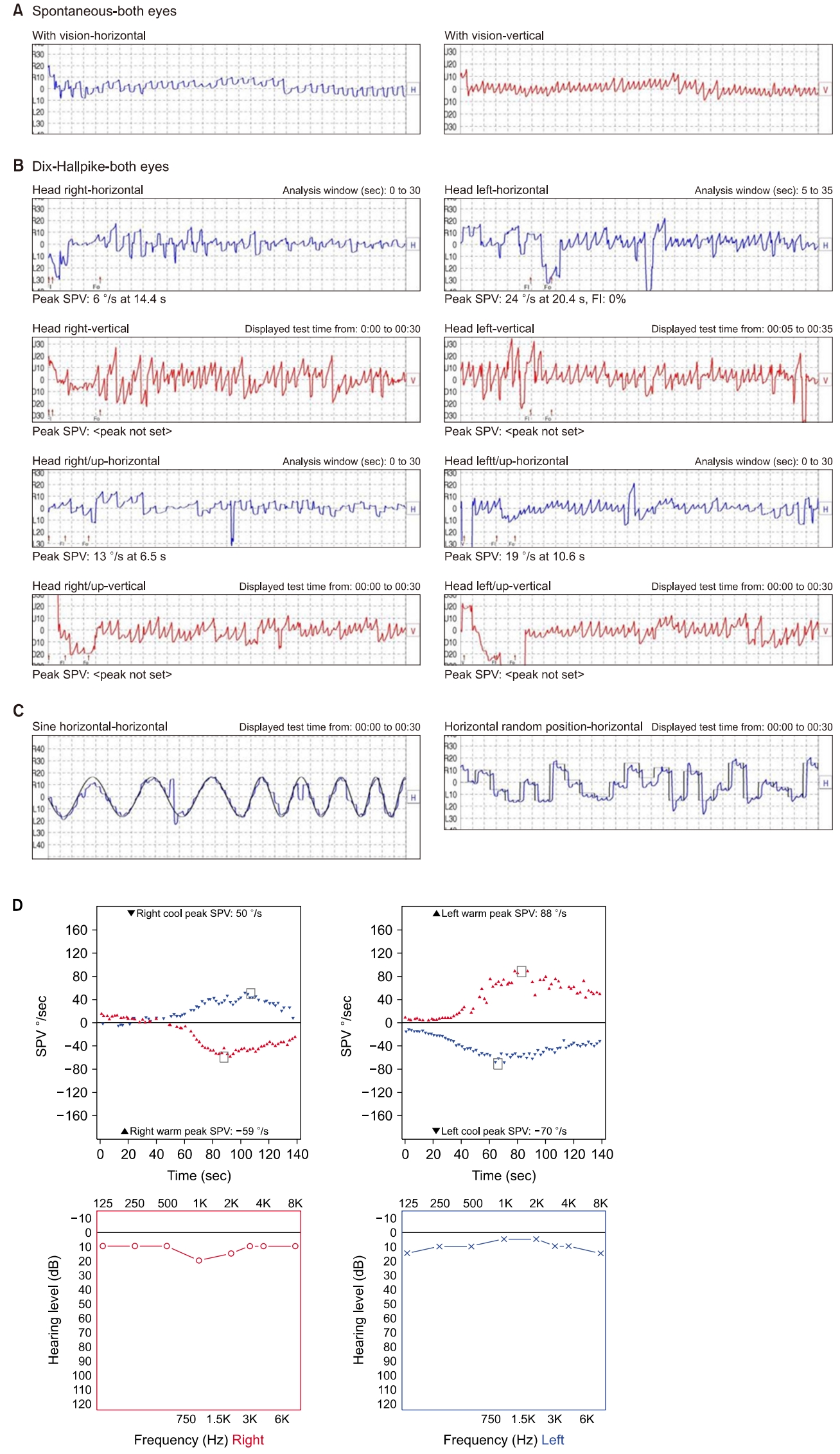
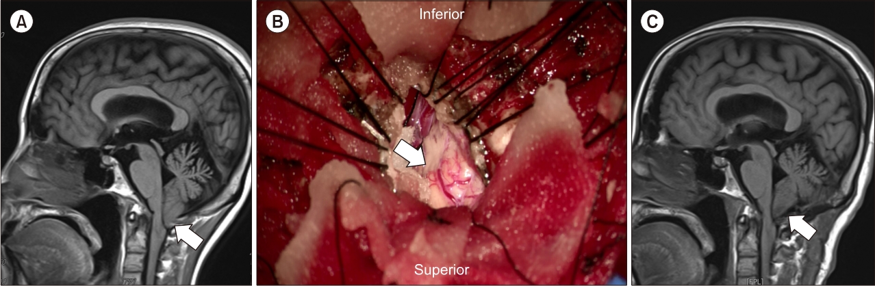
  • Arnold Chiari malformation is a disease which is characterized by herniation of a portion of the cerebellum through the foramen magnum. Symptoms vary depending on the extent of the affected area, including posterior neck pain, upper limb pain, paralysis, paresthesia, weakness, dizziness, and ataxia. Among the patients presenting with dizziness, nystagmus is frequently observed, which is primarily characterized by down-beating nystagmus. We experienced a 42- years-old female patient presented with vertigo and gait disturbance, who were diagnosed with type 1 Arnold-Chiari malformation and treated by surgical decompression.
아놀드-키아리 기형(Arnold-Chiari malformation)은 소뇌 또는 뇌간의 일부가 후두공(foramen magnum)을 통해 척추강으로 탈출되는 기형으로, 명확한 원인은 밝혀져 있지 않다[1]. 임상적으로는 탈출되어 압박된 소뇌편도로 인해 어지럼 및 실조가 생기면 어지럼 클리닉을 방문하는 경우가 있다. 자발성 하방안진은 대표적인 중추성 안진으로 소뇌 및 뇌간 부위에 병변이 있는 경우 관찰될 수 있는데, 아놀드-키아리 기형에서 대표적으로 관찰되는 안진이다[2]. 저자들은 자발성 하향안진 및 보행장애를 보이는 아놀드-키아리 기형 환자를 진단하고, 수술적 감압술로 치료한 증례를 문헌 고찰과 함께 보고하고자 한다.
42세 여자 환자가 수개월에 걸쳐 진행된 어지럼 및 보행장애를 주소로 이비인후과 외래로 내원하였다. 고혈압, 당뇨 등의 기저질환은 없었으며, 외상 및 이과 질환 치료의 과거력도 없었다.
환자는 간헐적으로 수 분간 악화되었다가 호전되는 회전성 어지럼을 호소하였는데, 체위 변경 시 악화되었고 휴식 시 완화되는 양상이었다. 이 외 이명 및 난청 등의 동반된 청각 증상은 없었다.
외래에서 시행한 비디오 프렌젤 안경 검사에서 자발성 하향안진이 관찰되었다. 주시유발에 의해 방향이 바뀌지 않았으나, 양측 Dix-Hallpike 검사 및 누워머리젖힘 head hanging 자세에서 심해지는 양상이었다(Supplementary Video 1). 또한, 롬버그검사 및 일자보행검사(tandem gait test)에서 흔들림을 보였다. 소뇌 기능 검사상, 양측 손가락코검사 및 발꿈치정강이검사에서 겨냥이상(dysmetria)을 보였고, 상반운동반복장애(dysdiadochokinesia)도 관찰되었다.
비디오 안진검사에서도 자발성 하향안진이 관찰되었다. 안진의 수직 성분은 격동 jerking 안진 형태였으나 속도 및 율동이 일정치 않았으며, 수평 성분은 사다리꼴에 가까운 진자운동성 안진을 보였고, 느린성분과 빠른성분이 명확히 구분되지 않는 형태였다. Dix-Hallpike 자세 및 누워머리젖힘 자세에서 수직 성분의 안진속도가 증가하였고, 시고정에도 억제되지 않았다(Fig. 1A, B).
안구운동검사상, 원활추종안구운동(smooth pursuit) 및 신속안구운동(saccade) 검사에서도 불규칙한 겨냥이상을 보였다(Fig. 1C). 또한, 온도안진검사(caloric test)에서 반고리관마비는 18%로 정상 범위였으나 시고정이 되지 않았다. 순음청력검사에서 양측 청력은 정상이었다(Fig. 1D).
소뇌 병변의 확인을 위해 뇌 자기공명영상을 시행하였다. 시상면 T1-가중 영상에서, 소뇌 편도가 대후두공으로부터 약 7 mm 하향 탈출을 보였고, 이로 인해 소뇌 및 뇌간 부위의 압박이 확인되었다. 확연한 척수공동증(syringomyelia)은 발견되지 않았다(Fig. 2A).
제1형 아놀드-키아리 기형으로 진단하여, 신경외과에서 후두 하 두개골 절제술을 통한 대후두공 감압술(foramen magnum decompression)을 시행하였다(Fig. 2B). 수술 후 2일째 시행한 자기공명영상에서 소뇌편도의 탈출과 압박은 호전되었다(Fig. 2C). 수술 후 2주 외래 방문 시, 어지럼과 실조 증상의 호전 추세 보였다. 수술 후 8개월째, 어지럼 증상은 없었고, 체위 변경 시에만 경미한 자발성 하향안진 만을 보였다. 롬버그검사 및 일자보행검사에서 흔들림은 보이지 않았다.
자발성 하향안진은 가장 흔한 중추성 안진 중 하나로, 상향안진과 달리 대부분 지속적이다. 대부분의 일상 생활 시 하향 주시를 하므로 상향안진에 비해 증상이 심한 경우가 많다[2]. 또한, 상향안진이 주로 뇌간의 국소 병변에 의해 발생하는 데 비해, 자발 하향안진은 원인이 밝혀지지 않는 경우도 많다. 확인된 유발 부위로는 주로 소뇌나 뇌간 부위의 국소 병변이 많고, 독성, 대사성 질환에 의한 경우도 있는데, Wagner 등[3]의 연구에서 총 117명의 하향 안진 환자들을 후향적으로 분석한 결과, 이 중 62%의 환자에서 원인 질환이 밝혀졌다. 또한, 전체 환자 중 6.8%가 아놀드-키아리 기형에 의한 것으로 보고하였다. 국내 연구에서도 자발성 하향안진으로 진단받은 환자 총 218명 중 원인이 밝혀진 것은 74.3%였으며, 원인은 크게 유전성, 혈관성, 염증성 순이었다. 이 연구에서 하향안진을 일으키는 병변 중 가장 많은 병소는 소뇌였으며, 4.1%의 환자에서 아놀드-키아리 기형이 원인 질환이었다.
하향안진의 발생 기전으로는 수직 방향의 전정안반사 이상, 원활추종안구운동의 장애, 이석기관-전정안반사 불균형 및 뇌간의 방정중 신경로 세포(paramedian tract cell)의 손상 등이 알려져있다[4-7]. 특히 소뇌 병변에서, 타래엽에서 기원하는 억제성 푸르킨예 세포(Purkinje cell)가 주로 상반고리관으로부터의 신호를 억제하므로 이 억제 기전에 문제가 생기면 하향안진이 발생한다고 알려져 있는데, 실제로 해당 부위를 절제할 때 하향안진이 발생하는 것을 확인한 바 있다[5,7-9]. 아놀드-키아리 기형에서 진행 정도에 따라 소뇌 압박이 심하지 않은 경우 체위 변화 시에만 하향안진이 확연해질 수 있는데, 이 경우 상반고리관 양성 돌발성 체위현훈과의 감별이 임상적으로 중요하겠다.
아놀드-키아리 기형은 두개골의 이상으로 소뇌 또는 뇌간이 후두공으로 탈출되는 선천성 질환으로 알려져 있다. 이 중 제1형은 소뇌편도의 후두공을 통한 5 mm 이상의 하향 탈출이 있는 경우로, 두개-척수 결합부의 골기형 또는 척수공동증 등이 동반될 수 있다[10]. 본 증례도 이에 해당하는 것으로, 뇌 자기공명영상검사로 소뇌편도의 탈출을 확인하고 진단하였다. 소아 때 증상이 나타나는 경우보다 30–50대에 이르러 증상이 발현되는 경우가 더 많다. 후두공 압박증후군(foramen magnum compression syndrome)이 동반되는 경우, 감각 저하, 소뇌 기능 이상 및 하부 뇌신경 마비(lower cranial nerve palsy) 등을 보일 수 있으며[10], 80%의 환자에서 안진, 복시 등의 안구 증상이 발생한다고 보고된 바 있다[11]. 아놀드-키아리 기형에서 발생하는 안진은 본 증례와 같이 하향안진이 대표적이나, 병의 진행 정도, 소뇌 및 뇌간 압박의 정도에 따라 여러 형태의 안진이 발생할 수 있겠다. 수평 안진, 주기교대안진(periodic alternating nystagmus), 눈모음안진(convergence nystagmus), 선천성안진(congenital nystagmus), 주시유발안진(gaze evoked nystagmus), 칼로릭-반동안진(rebound caloric nystagmus)과 같은 다양한 안진이 아놀드-키아리 기형에서 보고되었다[10-16]. 치료를 위해 수술적 감압술을 시행하고, 수술 이후 수개월에 걸쳐 안진과 원활추종안구운동이 호전된다고 보고되어 있는데, 병의 진행 정도에 따라 수술 직후 안진의 호전을 보인 경우도 있었다[10,17]. 보존적 치료를 시행한 제1형 아놀드-키아리 기형의 자연 경과는 매우 다양한데, 147명의 환자를 4.6년간 추적 관찰한 연구에서는 14명의 환자가 추적 중 증상이 악화되어 수술적 치료를 받은 것으로 보고하였다[18].
본 증례와 같이 자발성 햐향안진이 관찰된 경우, 즉시 소뇌기능을 평가하고 적절한 영상검사를 통해 아놀드-키아리 기형을 감별 진단하여 적절한 치료를 시행하는 것이 임상적으로 중요하겠다.

이해관계(CONFLICT OF INTEREST)

저자들은 이 논문과 관련하여 이해관계의 충돌이 없음을 명시합니다.

연구 지원(FUNDING/SUPPORT)

이 연구는 2017년도 정부(교육과학기술부)의 재원으로 한국연구재단의 지원을 받아 수행된 기초연구사업입니다(NRF-2017R1D1A3B03030968).

저자 기여(AUTHOR CONTRIBUTIONS)

Conceptualization, Funding acquisition, Investigation, Visualization: MK; Data curation, Formal analysis, Methodology: MK, YJC; Writing–Original Draft: MK; Writing–Review & Editing: MK, YJC.

All authors read and approved the final manuscript.

Supplementary Video 1 can be found via https://doi.org/10.21790/rvs.2023.22.4.132.
Fig. 1.
The result of audio-vestibular tests. (A) Spontaneous down-beating nystagmus on videonystgmography. (B) Nystagmus at bilateral Dix-Hallpike test. (C) Smooth pursuit and saccade test. (D) Caloric test and pure tone audiometry. SPV, slow phase velocity.
rvs-22-4-132f1.jpg
Fig. 2.
Magnetic resonance (MR) images before and after surgical decompression. (A) Herniated cerebellar tonsil (arrow) on the preoperative T1-weighted MR images. (B) Herniated cerebellar tonsil (arrow) during surgical decompression. (C) Decompressed cerebellar tonsil (arrow) on the postoperative MR images.
rvs-22-4-132f2.jpg
  • 1. Russell GE, Wick B, Tang RA. Arnold-Chiari malformation. Optom Vis Sci 1992;69:242–7.ArticlePubMed
  • 2. Kim KT, Jung I, Kim JS. Clinical characteristics of spontaneous downbeat nystagmus in Koreans: analysis of 218 cases. J Korean Neurol Assoc 2016;34:27–32.ArticlePDF
  • 3. Wagner JN, Glaser M, Brandt T, Strupp M. Downbeat nystagmus: aetiology and comorbidity in 117 patients. J Neurol Neurosurg Psychiatry 2008;79:672–7.ArticlePubMed
  • 4. Baloh RW, Spooner JW. Downbeat nystagmus: a type of central vestibular nystagmus. Neurology 1981;31:304–10.ArticlePubMed
  • 5. Zee DS, Friendlich AR, Robinson DA. The mechanism of downbeat nystagmus. Arch Neurol 1974;30:227–37.ArticlePubMed
  • 6. Halmagyi GM, Rudge P, Gresty MA, Sanders MD. ownbeating nystagmus. A review of 62 cases. Arch Neurol 1983;40:777–84.ArticlePubMed
  • 7. Nakamagoe K, Iwamoto Y, Yoshida K. Evidence for brainstem structures participating in oculomotor integration. Science 2000;288:857–9.ArticlePubMed
  • 8. Marti S, Straumann D, Glasauer S. The origin of downbeat nystagmus: an asymmetry in the distribution of on-directions of vertical gaze-velocity Purkinje cells. Ann N Y Acad Sci 2005;1039:548–53.ArticlePubMed
  • 9. Böhmer A, Straumann D. Pathomechanism of mammalian downbeat nystagmus due to cerebellar lesion: a simple hypothesis. Neurosci Lett 1998;250:127–30.ArticlePubMed
  • 10. Korres S, Balatsouras DG, Zournas C, Economou C, Gatsonis SD, Adamopoulos G. Periodic alternating nystagmus associated with Arnold-Chiari malformation. J Laryngol Otol 2001;115:1001–4.ArticlePubMed
  • 11. Paul KS, Lye RH, Strang FA, Dutton J. Arnold-Chiari malformation. Review of 71 cases. J Neurosurg 1983;58:183–7.PubMed
  • 12. Spooner JW, Baloh RW. Arnold-Chiari malformation: improvement in eye movements after surgical treatment. Brain 1981;104:51–60.ArticlePubMed
  • 13. Mossman SS, Bronstein AM, Gresty MA, Kendall B, Rudge P. Convergence nystagmus associated with Arnold-Chiari malformation. Arch Neurol 1990;47:357–9.ArticlePubMed
  • 14. Nakahara H, Murofushi T. Congenital-type nystagmus in Arnold-Chiari malformation. Otolaryngol Head Neck Surg 2003;128:598–600.ArticlePubMedPDF
  • 15. Ghasia FF, Gulati D, Westbrook EL, Shaikh AG. Viewing condition dependence of the gaze-evoked nystagmus in Arnold Chiari type 1 malformation. J Neurol Sci 2014;339:134–9.ArticlePubMed
  • 16. Kumar A, Pieri A, Krol G. Rebound caloric nystagmus. Laryngoscope 1993;103:1205–13.ArticlePubMed
  • 17. Pedersen RA, Troost BT, Abel LA, Zorub D. Intermittent downbeat nystagmus and oscillopsia reversed by suboccipital craniectomy. Neurology 1980;30:1239–42.ArticlePubMed
  • 18. Strahle J, Muraszko KM, Kapurch J, Bapuraj JR, Garton HJ, Maher CO. Natural history of Chiari malformation Type I following decision for conservative treatment. J Neurosurg Pediatr 2011;8:214–21.ArticlePubMed

Figure & Data

References

    Citations

    Citations to this article as recorded by  

      • PubReader PubReader
      • ePub LinkePub Link
      • Cite
        CITE
        export Copy
        Close
        Download Citation
        Download a citation file in RIS format that can be imported by all major citation management software, including EndNote, ProCite, RefWorks, and Reference Manager.

        Format:
        • RIS — For EndNote, ProCite, RefWorks, and most other reference management software
        • BibTeX — For JabRef, BibDesk, and other BibTeX-specific software
        Include:
        • Citation for the content below
        Arnold-Chiari Malformation Presented with Spontaneous Down-Beating Nystagmus and Gait Disturbance
        Res Vestib Sci. 2023;22(4):132-136.   Published online December 15, 2023
        Close
      • XML DownloadXML Download
      Figure
      We recommend
      Related articles

      Res Vestib Sci : Research in Vestibular Science