Skip Navigation
Skip to contents

Res Vestib Sci : Research in Vestibular Science

OPEN ACCESS
SEARCH
Search

Articles

Page Path
HOME > Res Vestib Sci > Volume 23(1); 2024 > Article
Case Report
A case of cerebellopontine angle meningioma presenting as neurovascular compression syndrome of the 8th cranial nerve
Jeongin Jang1orcid, Sung Kwang Hong1,2orcid, Joonho Song3orcid, Hyung-Jong Kim1orcid, Hyo-Jeong Lee1,2orcid
Clinical and Experimental Otorhinolaryngology 2024;23(1):28-31.
DOI: https://doi.org/10.21790/rvs.2023.141
Published online: March 15, 2024

1Department of Otorhinolaryngology-Head and Neck Surgery, Hallym University College of Medicine, Chuncheon, Korea

2Laboratory of Brain & Cognitive Sciences for Convergence Medicine, Hallym University College of Medicine, Chuncheon, Korea

3Department of Neurosurgery, Hallym University College of Medicine, Chuncheon, Korea

Corresponding Author: Hyo-Jeong Lee Department of Otolaryngology, Hallym University Medical Center, 22 Gwanpyeong-ro 170beon-gil, Dongan-gu, Anyang, 14068, Korea E-mail: hyojlee@hallym.ac.kr
• Received: November 3, 2023   • Revised: November 23, 2023   • Accepted: November 24, 2023

© 2024 The Korean Balance Society

This is an open access article distributed under the terms of the Creative Commons Attribution Non-Commercial License (http://creativecommons.org/licenses/by-nc/4.0) which permits unrestricted non-commercial use, distribution, and reproduction in any medium, provided the original work is properly cited.

  • 227 Views
  • 2 Download
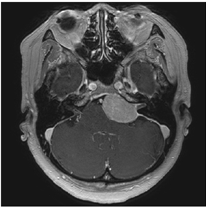
  • A 54-year-old female patient presented with paroxysmal tinnitus and vertigo for 2 years, which have repeatedly occurred while rotating her neck, and lasted about 10 seconds. An anticonvulsant medication was prescribed with a diagnosis of microvascular compression syndrome on the 8th cranial nerve and audiovestibular evaluation and magnetic resonance imaging (MRI) scan were appointed a week later. In her next visit, she reported the disappearance of paroxysmal audiovestibular symptoms after medication. In the left ear, canal paresis and abnormal auditory brainstem response were observed. In MRI, a large meningioma in the cerebellopontine angle in the vicinity of the internal auditory canal orifice was detected, that was surgically resected by a neurosurgeon. After surgical removal of the tumor, she reported continuous dizziness due to vestibular nerve injury, but the paroxysmal attack of tinnitus and vertigo disappeared without anticonvulsant medication. This case suggests that an imaging study is mandatory when diagnosing microvascular compression syndrome on the 8th cranial nerve.
Neurovascular compression syndrome (NVCS) of the 8th cranial nerve typically presents with paroxysmal audiovestibular symptoms due to vascular compression of the nerve at the central segment lying in the cerebellopontine angle (CPA). This disorder can cause various combinations of auditory and vestibular symptoms, which have been called different disease categories, such as vestibular paroxysmia (VP) [1,2], disabling positional vertigo [3], typewriter tinnitus [4,5], and cochleovestibular compression syndrome (CVCS) [6]. Diagnostic criteria for CVCS and VP have been reported and revised recently, all of them defining CVCS and VP in terms of clinical symptoms and response to antiepileptic medications [1,2,4,5]. In this case report, we present a case of CPA meningioma presenting as NVCS of the 8th cranial nerve and discuss the value of laboratory tests.
This study has been approved by the Institutional Review Board of the Hallym University Sacred Heart Hospital (No. 2017-I059).
A 54-year-old woman presented to our clinic with a primary complaint of repeated tinnitus with spinning vertigo of 2 years’ duration. The paroxysmal audiovestibular symptoms lasted approximately 10 seconds and were triggered when she rapidly rotated her head laterally. She also had decreased hearing and aural fullness in her left ear. She showed spontaneous nystagmus and head-shaking nystagmus with a fast phase beating to the right during examination with Frenzel goggles. Audiovestibular laboratory tests and magnetic resonance imaging (MRI) of the internal auditory canal were scheduled for 1 week later and anticonvulsant medication (oral oxcarbazepine, 300 mg/day) was prescribed. She returned to the clinic after all the laboratory tests were completed. She reported that the paroxysmal dizziness and tinnitus had ceased, but once when she missed a dose of medication, the symptoms reappeared. She had a normal audiogram with 100% word recognition scores in both ears. Caloric testing showed 52% canal paresis in the left ear. Auditory brainstem response (ABR) testing showed an abnormal waveform on the left side (Fig. 1). MRI revealed a well-enhanced, 3.6 cm broad-based mass in the left CPA (Fig. 2). She underwent neurosurgical excision as well as postoperative gamma knife radiotherapy for the residual mass. Subsequent histopathologic examination confirmed a meningothelial meningioma. The patient discontinued anticonvulsant medication after surgery, and the paroxysmal attacks of dizziness and tinnitus resolved, though she had some continuous dizziness that improved with vestibular rehabilitation exercises.
NVCS of the 8th cranial nerve is characterized by repeated brief attacks of vestibulocochlear symptoms. It typically improves dramatically when treated with low-dose anticonvulsant medication, but microvascular decompression surgery is indicated for medically intractable cases [7,8]. Audiometry and vestibular function tests are usually recommended, as many patients exhibit signs of mild-to-moderate unilateral hypofunction during attack-free intervals, which may be helpful in determining the affected side [3,4]. ABR and MRI have been suggested as diagnostic tests for NVCS but both have questionable diagnostic efficacy. In a previous case series, pathologic ABR changes were found in less than one-third of cases [2]. Neurovascular contact demonstrated by MRI is only suggestive and does not definitively diagnose NVCS even with a high-tesla (7T) experimental machine [5].
The roles of both ABR testing and MRI in identifying NVCS need further evaluation. However, we suggest performing either ABR testing or MRI when evaluating patients with suspected NVCS of the 8th cranial nerve to exclude mass lesions in the CPA, which can be fatal. In this case, although the clinical symptoms and response to anti-epileptic treatment fulfilled the diagnostic criteria for VP, abnormalities found through ABR testing and MRI led to a different final diagnosis. The CPA meningioma stereotypically leads to symptoms relating to mass effect on cranial nerves III, V, VII, and VIII, while in NVCS syndrome, audiovestibular symptoms arise from ephaptic transmission between demyelinated axons by the pulsatile compression of the responsible vessel. Thus, there could be differences in symptom presentation between the two disease categories. However, the symptoms related to the compression on the 8th cranial nerve would be similar, whatever the compressing tissue is. We speculated that the mass effect on the 8th cranial nerve could also lead to audiovestibular symptoms mimicking those in NVCS. Still, the conclusion about pathomechanism should be substantiated with further study.

Funding/Support

This work was supported by the National Research Foundation of Korea (NRF) grant funded by the Korean Government (2022R1A2C1004862).

Conflicts of Interest

No potential conflict of interest relevant to this article was reported.

Availability of Data and Materials

All data generated or analyzed during this study are included in this published article. For other data, these may be requested through the corresponding author.

Authors' Contributions

Conceptualization, Resources: HJL, JS; Data curation: JJ; Formal analysis: JJ, SKH; Funding acquisition, Project administration, Supervision, Validation: HJL; Investigation: SKH; Methodology: HJL, HJK; Writing–original draft: HJL, JJ; Writing–review & editing: all authors.

All authors read and approved the final manuscript.

Fig. 1.
Results from caloric test and auditory brainstem response (ABR). The caloric test revealed 52% canal paresis in the left ear. The peak latency in ABR was marked for each peak I and V but the remaining peaks (peak II, III, and IV) were not detected on the left side, while I–V peaks clearly were observed on the right side. SCV, slow component velocity; FI, fixation index; F, fixation; NF, none-fixation; RVR, relative vestibular reduction; UW, unilateral weakness; DP, directional preponderance.
rvs-2023-141f1.jpg
Fig. 2.
Gadolinium-enhanced magnetic resonance imaging of the internal auditory canal. A well-enhancing broad-based mass was examined in the left cerebellopontine angle region.
rvs-2023-141f2.jpg
  • 1. Hüfner K, Barresi D, Glaser M, et al. Vestibular paroxysmia: diagnostic features and medical treatment. Neurology 2008;71:1006–1014.ArticlePubMed
  • 2. Strupp M, Lopez-Escamez JA, Kim JS, et al. Vestibular paroxysmia: diagnostic criteria. J Vestib Res 2016;26:409–415.ArticlePubMedPMC
  • 3. Jannetta PJ, Møller MB, Møller AR. Disabling positional vertigo. N Engl J Med 1984;310:1700–1705.ArticlePubMed
  • 4. Koo YJ, Kim HJ, Choi JY, Kim JS. Vestibular paroxysmia associated with typewriter tinnitus: a case report and literature review. J Neurol 2021;268:2267–2272.ArticlePubMedPDF
  • 5. Levine RA. Typewriter tinnitus: a carbamazepine-responsive syndrome related to auditory nerve vascular compression. ORL J Otorhinolaryngol Relat Spec 2006;68:43–47.ArticlePubMedPDF
  • 6. De Ridder D, Vanneste S, Adriaensens I, Lee AP, van de Heyning P, Möller A. Vascular compression of the cochlear nerve and tinnitus: a pathophysiological investigation. Acta Neurochir (Wien) 2012;154:807–813.ArticlePubMedPDF
  • 7. De Ridder D, Heijneman K, Haarman B, van der Loo E. Tinnitus in vascular conflict of the eighth cranial nerve: a surgical pathophysiological approach to ABR changes. Prog Brain Res 2007;166:401–411.PubMed
  • 8. Shandal V, Fernández-Conejero I, Sekula R, Crammond D, Thirumala P, Anetakis K, et al. Microvascular decompression for cranial nerve disorders. In: Seubert CN, Balzer JR, editors. Koht, Sloan, Toleikis’s monitoring the nervous system for anesthesiologists and other health care professionals. 3rd ed. Springer, Cham; 2022. p. 465-493.

Figure & Data

References

    Citations

    Citations to this article as recorded by  

      • PubReader PubReader
      • ePub LinkePub Link
      • Cite
        CITE
        export Copy
        Close
        Download Citation
        Download a citation file in RIS format that can be imported by all major citation management software, including EndNote, ProCite, RefWorks, and Reference Manager.

        Format:
        • RIS — For EndNote, ProCite, RefWorks, and most other reference management software
        • BibTeX — For JabRef, BibDesk, and other BibTeX-specific software
        Include:
        • Citation for the content below
        A case of cerebellopontine angle meningioma presenting as neurovascular compression syndrome of the 8th cranial nerve
        Res Vestib Sci. 2024;23(1):28-31.   Published online March 15, 2024
        Close
      • XML DownloadXML Download
      Figure
      We recommend
      Related articles

      Res Vestib Sci : Research in Vestibular Science