Abstract
-
Objectives
- The objective of this study was to analyze vestibulocochlear function results in patients identified with isolated semicircular canal (SCC) hypofunction using the video head impulse test (vHIT).
-
Methods
- A retrospective review was conducted on the clinical records of 123 patients diagnosed with isolated SCC hypofunction based on vHIT results. Among these patients, 72 had isolated posterior SCC (PSCC) hypofunction, 25 had isolated lateral SCC (LSCC) hypofunction, and 26 had isolated anterior SCC (ASCC) hypofunction. Descriptive analyses were performed on various vestibulocochlear tests including pure tone audiometry, sinusoidal harmonic acceleration (SHA), spontaneous nystagmus (SN), head-shaking nystagmus (HSN), caloric testing, and cervical vestibular evoked myogenic potential, with results analyzed separately for each SCC hypofunction group.
-
Results
- The study found that 66.0% of the evaluated patients exhibited abnormal results in at least one vestibulocochlear function test. PSCC hypofunction patients showed a significantly higher incidence of hearing loss compared to ASCC and LSCC hypofunction patients. LSCC hypofunction patients exhibited higher rates of corrective saccade, phase asymmetry of SHA, and SN abnormalities compared to other SCC hypofunction patients. Additionally, the rates of corrective saccade and phase asymmetry of SHA were also higher in LSCC hypofunction patients. ASCC hypofunction patients demonstrated significantly higher rates of normal corrective saccade, phase lead of SHA, and SN.
-
Conclusions
- The analysis of this study suggests that even in cases where vHIT indicates isolated SCC hypofunction, additional vestibulocochlear function tests should be conducted to identify any associated vestibulocochlear dysfunctions. This highlights the importance of comprehensive evaluation to accurately diagnose and manage patients with SCC hypofunction.
-
Keywords: Video head impulse test; Semicircular canal; Vestibular function tests; Nystagmus
-
중심단어: 비디오두부충동검사, 세반고리관, 전정기능검사, 안진
서 론
두부충동 검사(head impulse test, HIT)는 전정안반사(vestibulo-ocular reflex, VOR)를 이용하여 말초 전정기능의 이상을 측정하는 검사이다[1]. VOR은 머리의 움직임과 같은 속도의 반대 방향으로 눈을 움직여 물체의 상을 망막에 맺게 하는 반사운동으로, HIT 시 검사자는 환자에게 고글을 씌운 뒤 머리를 빠르게 움직이게 하여 VOR을 유도한다. 그러나 전정기능에 문제가 있는 경우 머리를 돌릴 때 환자의 눈이 머리 움직임의 속도에 맞추어 움직이지 못하여 망막에 맺히는 주시점의 상이 흔들리게 되고, 이 때 주시점 상을 맺기 위한 보상적 교정단속운동(corrective saccade)을 하여 안구가 놓쳤던 목표물을 볼 수 있게 된다. 최근에는 비디오 HIT (video HIT, vHIT) 기기의 도입으로 머리의 움직임에 따른 눈의 움직임의 정도를 이득(gain)으로 계산하고 주시상을 맺기 위한 교정단속운동을 정량적으로 측정하여 전정기능의 이상 유무를 판정할 수 있다[2].
전정기능 평가 시 VOR를 이용하는 검사로는 온도안진검사(caloric test), 회전의자 검사(rotary chair test)가 있다. 온도안진검사는 0.002–0.004 Hz의 저주파의 머리의 움직임을, 회전의자 검사는 0.01–0.64 Hz의 중주파의 머리의 움직임을 반영한다[3]. vHIT 는 약 0.1–10 Hz의 비교적 빠른 머리의 움직임을 반영하여 일상생활의 움직임에 대한 VOR을 볼 수 있다. 온도안진검사는 양측의 전정기관을 개별적으로 자극하는 장점이 있지만, 양측성 병변에 민감하지 않고 수평반고리관의 기능만을 제한적으로 볼 수 있다. 또한 온도자극을 위하여 외이도에 공기 혹은 물을 주입하고 어지러움을 유발하기 때문에 환자가 불쾌감을 느낄 수 있다는 단점이 있다[4]. 회전의자 검사는 온도안진검사를 실시할 수 없는 환자, 예를 들어 외이와 중이에 병변이 있는 환자, 소아 등을 검사할 수 있다는 장점이 있지만, 검사 기기가 고가이며 장소에 제약이 있다는 단점이 있고[5], 온도안진검사와 마찬가지로 수평반고리관의 기능을 주로 본다.
전정기능 저하의 진단에 기존에 이용하였던 공막탐색 코일 HIT와 진단율에 차이가 없는 vHIT의 임상적 의의와 진단의 정확성, 그리고 질환별 차이에 대한 연구자들의 관심이 최근 늘어나고 있다[2]. 또한 기존의 전정기능 검사와 달리 6개의 기능을 각각 객관적으로 살펴볼 수 있다는 점이 vHIT의 가장 큰 특징이자 임상적 유용성이라고 할 수 있다[6-9]. vHIT로는 다른 전정기능 평가에서 가능하지 않은 한 개의 반고리관 이상도 감지할 수 있다. 임상적으로 한 개의 반고리관에만 이상이 있는 경우는 흔하지는 않지만 규칙적으로 발생할 수 있으며[10], 한 개의 반고리관만 이상이 있을 경우 어지럼이나 현훈 이외에 다른 증상이 나타나지 않아 진단하기 힘들 수 있다. 따라서 임상에서는 vHIT 결과 한 개의 반고리관에서만 이상을 보인 환자들을 대상으로 임상적 특징과 진단 결과를 더욱 세분화한다면 전정 재활에 있어서 효과적일 것이다[11].
본 연구에서는 vHIT에서 각 반고리관 한 개에서만 기능 저하를 보인 대상자를 후향적으로 선별하여 그 환자들의 전정와우 기능의 패턴을 분석하고 비교하고자 한다. 본 연구의 결과는 vHIT에 대한 이해를 높이는 동시에 추후 전정기능 진단을 위한 자료로 사용할 수 있을 것이다.
대상 및 방법
1. 연구 대상
2019년 1월부터 2021년 6월까지 2년 6개월 동안 단국대학교병원 이비인후과에 어지럼증으로 내원한 환자 중 vHIT를 수직반고리관과 수평반고리관 모두 시행한 대상자 1,145명을 분석하였다. 대상자는 어지럼으로 처음 내원 시 시행한 vHIT를 결과로 분리하였으며, 그 외 전정와우 기능을 보기 위한 순음청력검사, 회전의자 검사 중 정현파 회전검사(sinusoidal harmonic acceleration), 냉온교대온도안진검사, 경부전정유발근전위(cervical vestibular evoked myogenic potential, cVEMP) 검사, 자발 안진검사(spontaneous nystagmus), 두진 후 안진검사(head-shaking nystagmus)는 vHIT 검사일 기준 14일 전후로 검사한 결과 중 가장 가까운 검사일을 기준으로 후향적으로 분석하였다. 모든 검사는 5년 이상의 청각 및 전정 평가 이력을 가진 검사자 3명이 검사를 실시하였다. 이 연구는 단국대학교병원 기관윤리위원회의 승인을 받은 후 진행하였으며(No. 2021-12-012-002), 후향적 연구 특성상 환자 동의서는 면제되었다.
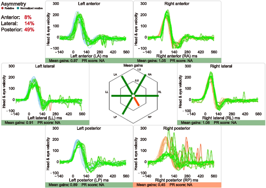
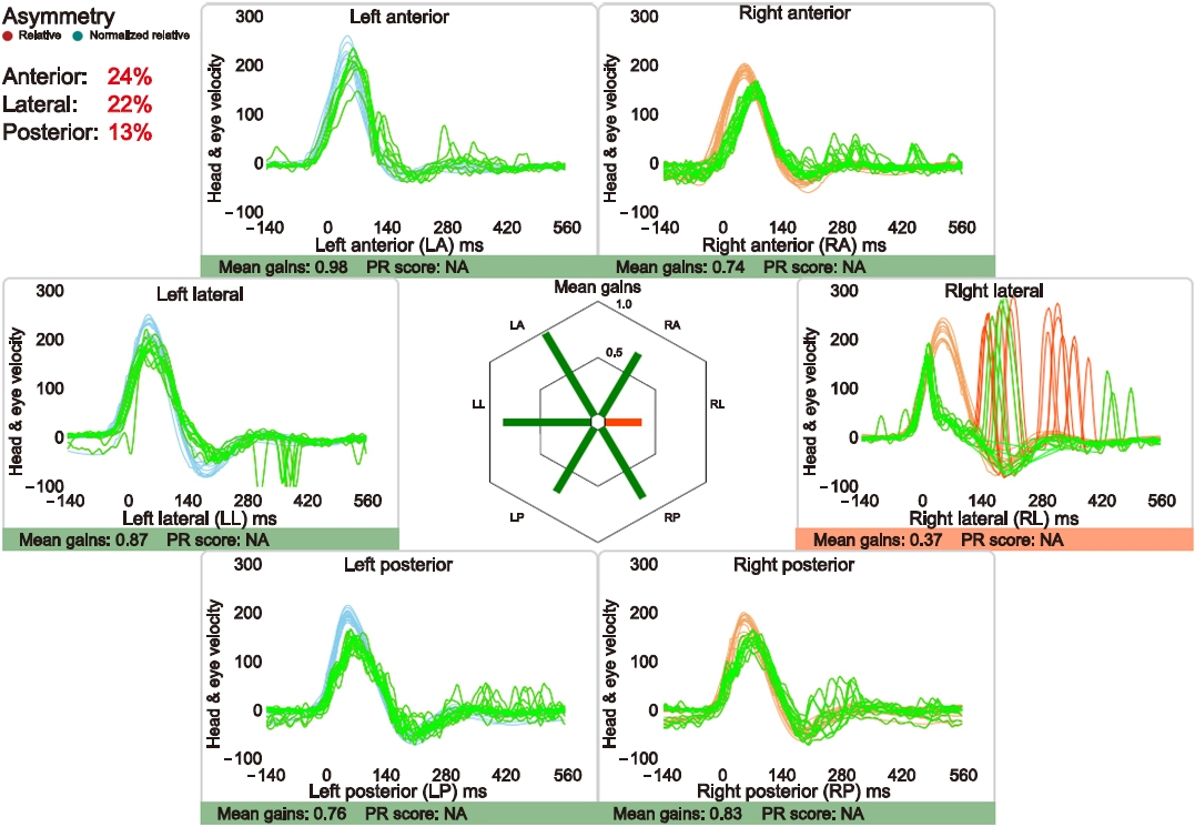
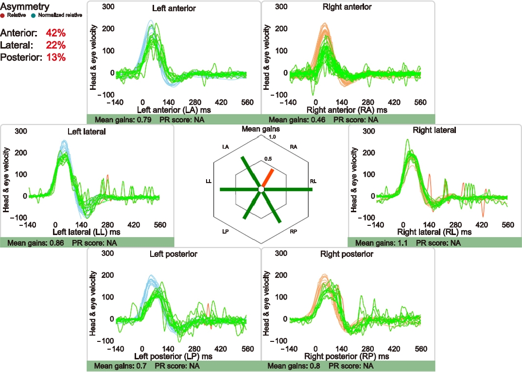
vHIT 결과에서 이득 저하(수평반고리관 <0.8, 수직반고리관 <0.7)를 보인 한 개의 반고리관 외 다른 다섯 개의 반고리관의 이득이 정상일 때 unilateral isolated semicircular canal (SCC) hypofunction이라고 정하였다[12]. vHIT 검사에서 unilateral isolated SCC hypofunction의 예를 Figs. 1–3에 제시하였다. 총 1,145명 중에서 unilateral isolated SCC hypofunction을 보인 사람은 123명이며, 이 중에서 unilateral isolated posterior SCC (PSCC) hypofunction은 72명(여자 41명, 남자 31명, 평균 나이 53.9±14.2세), unilateral isolated lateral SCC (LSCC) hypofunction은 25명(여자 15명, 남자 10명, 평균 나이 49.2±14.6세), unilateral isolated anterior SCC (ASCC) hypofunction은 26명(여자 22명, 남자 4명, 평균 나이 49.5±15.4세)이다.
vHIT에서 unilateral isolated SCC hypofunction을 보인 대상자 123명 중 순음청력검사, 정현파 회전검사, 냉온교대온도안진검사, cVEMP, 자발 안진검사, 두진 후 안진검사를 시행한 결과는 Table 1과 같다.
2. 검사 방법
1) vHIT
vHIT (ICS impulse, GN Otometrics) 시행 시 환자에게 비디오 고글을 착용케 하고 검사하였다. 머리 회전각도는 10°–20°이며, 최대 각속도는 100°–250°/초로 머리를 회전시켰다. 검사는 반고리관별로 최소 10회씩 반복하였으며 vHIT 결과 이득이 수평반고리관 <0.8, 수직반고리관 <0.7로 떨어졌을 경우 양성으로 판정하였다[12]. 단, unilateral isolated SCC hypofunction의 경우 양 귀 6개의 반고리관 중에서 기능 저하를 보인 한 개의 반고리관 외 다른 5개의 반고리관은 이득이 정상이어야 한다.
2) 순음청력검사
순음청력검사(Otometrics Madsen Astera, Otometrics) 시 주파수 250, 500, 1,000, 2,000, 4,000 Hz에서의 역치를 분석하였다. 한 주파수 이상 비대칭적으로 양이 청력 역치 차가 15 dB HL 이상 차이가 발생할 경우 청력 저하가 있다고 판단하였으며[13], 역치가 더 상승한 측을 확인하였다. 단, 전음성 또는 혼합성 난청을 보인 대상자는 제외하였다.
3) 정현파 회전검사
회전의자 검사(System 2000, Micromedical Technologies Inc.)에서 정현파 회전검사는 60°/sec2 의 최대 각속도로 진행하였으며, 회전 주파수는 0.01, 0.02, 0.04, 0.08, 0.16, 0.32, 0.64 Hz 순으로 진행하였다. 회전의자 검사를 시행한 41명의 환자 중 12명은 0.02, 0.08, 0.32 Hz를 시행하지 않았다. 검사가 시행된 주파수 중 이득이 한 개 이상 정상 기준치보다 낮게 나온 경우, 위상 차 선행(phase lead)을 보인 경우, 또는 위상 비대칭이 15% 이상으로 나타난 경우 모두 양성으로 판정하고 병변 방향을 확인하였다.
4) 냉온교대온도안진검사
냉온교대온도안진검사(VISUALEYES Spectrum, Micromedical Technologies Inc.)는 환자가 비디오 고글을 착용 후 머리를 30°정도 든 자세를 유지할 수 있도록 베개를 베고 누워 안정을 취한 후에 검사를 진행하였다. 물의 온도는 각각 30 ℃의 찬물과 44 ℃의 따뜻한 물을 양측 귀에 교대로 주입하여 나타나는 안진을 기록하여 분석하였다. 반고리관 마비(canal paresis) 값은 안진의 느린 성분 속도의 최대값을 이용한 Jonkee’s 공식을 통해 확인하였으며[14], 값이 25%를 초과한 경우 양성으로 판단하였다.
5) 경부전정유발근전위 검사
환자를 방음실에 있는 침대에 눕혀 편안한 상태를 유지하도록 한 후 검사를 진행하였다. 전극을 이마와 흉쇄유돌근(sternocleido mastoid muscle)에 붙인 후, 검사 귀의 반대 방향으로 고개를 돌리고 머리를 바닥으로부터 올려 최대한 근의 긴장도를 높인 후에 cVEMP (Bio-logic Navigator PRO, Natus)검사를 시행하였다. 검사 자극음은 95 dB nHL의 500 Hz 톤버스트음을 사용하였다. 검사 결과 파형이 관찰되지 않거나 양이 간 진폭 차 비(interaural amplitude difference ratio, IAD)가 40% 이상인 경우를 양성으로 정의하였다. IAD는 rectification하여 계산하였다[15].
6) 자발 안진검사 및 두진 후 안진 검사
비디오 안진검사(System 2000)는 환자의 눈의 움직임을 기록하여 측정하는 검사로, 자발 안진검사와 두진 후 안진검사를 시행하여 나타나는 안구의 움직임을 확인하였다. 자발 안진은 앉아있는 상태에서 비디오 고글을 씌운 후 눈의 움직임을 확인하였다. 두진 후 안진검사는 비디오 고글을 착용한 상태에서 환자의 고개를 30° 앞으로 숙이게 한 후에 좌우로 20–30회, 2 Hz의 빈도로 고개를 흔든 다음 안진의 유무와 방향을 확인하였다. 안진의 방향은 빠른 성분의 방향으로 정하였다.
3. 통계 분석
통계 프로그램은 IBM SPSS Statistics ver. 25.0 (IBM Corp.)을 사용하여 분석하였다. 각 반고리관에 따라 전정기능검사들을 양성과 음성으로 나눠 비모수 검정인 카이제곱검정(chi-square test)을 통해 각 반고리관별 차이를 분석하였으나, 전정와우 기능 검사 중에서 표본 크기가 작은 경우 Fisher의 정확검정을 통해 유의 확률을 확인하였다. 유의 확률은 p<0.05인 경우 통계적으로 유의한 차이가 있는 것으로 판정하였다.
결 과
vHIT를 진행한 환자 1,145명 중에서 unilateral isolated SCC hypofunction을 보인 사람은 123명이었으며, 이 중 unilateral isolated PSCC hypofunction 환자는 72명, unilateral isolated LSCC hypofunction 환자는 25명, unilateral isolated ASCC hypofunction 환자는 26명이었다. 본 연구에서 이득 저하와 교정단속운동을 같이 보이는 정도는 unilateral isolated PSCC hypofunction 환자는 72명 중 46명(63.9%), unilateral isolated LSCC hypofunction 환자는 25명중에서 22명(88.0%), unilateral isolated ASCC hypofunction 환자는 26명중에서 1명(3.8%)이었다(p<0.05). vHIT에서 100°/초 이상의 진폭이 반복적으로 50% 이상 나타났을 경우에 교정 단속운동을 보였다고 정하였다[16,17].
1. Unilateral isolated SCC hypofunction를 보인 환자들의 각 전정와우 검사별 결과
1) 순음청력검사 결과
순음청력검사는 양 귀 청력의 차이가 15 dB HL 이상 차이가 나는 경우를 비대칭한 청력이라고 판단하였다. Unilateral isolated PSCC hypofunction를 보인 환자 중에서 순음청력검사 결과 한 개의 주파수 이상 비대칭적으로 청력 저하를 보인 사람은 45명 중에 28명(62.2%)이었으며, 이 중 vHIT에서 기능 저하를 보인 방향과 일치하는 환자는 27명(96.4%)이었다(Table 2). 이 중 전 주파수 영역이 모두 저하한 경우는 12명, 주로 고주파 대역 이상에서 청력이 저하한 경우가 12명, 저주파수 대역이 저하한 경우가 3명이었다. Unilateral isolated LSCC hypofunction를 보인 환자 중 청력 저하를 보인 환자는 15명 중에서 7명(46.7%)이었으며, 이 중 병변 방향이 일치한 사람은 4명(57.1%)였다(Table 3). Unilateral isolated ASCC hypofunction을 보인 환자 중에는 청력 저하가 17명 중 7명(41.2%)이며, 이 중 병변 방향이 일치한 사람은 3명(42.9%)이었다(Table 4). Unilateral isolated SCC hypofunction을 보인 후반고리관, 측반고리관, 상반고리관에 따라 청력 저하를 동반한 경우는 서로 무의미한 차이를 보였으나, 청력 저하를 보인 환자 중 청력 저하의 방향과 vHIT의 이득 저하 방향이 동일한 경우는 unilateral isolated LSCC hypofunction과 unilateral isolated ASCC hypofunction에 비해 unilateral isolated PSCC hypofunction에서 유의미하게 방향 일치성이 높았다(p<0.05).
2) 정현파 회전검사 결과
Unilateral isolated PSCC hypofunction을 보인 환자 26명 중 이득 저하를 보인 사람은 11명(42.3%), 위상 차 선행을 보인 사람은 4명(15.4%), 좌우 비대칭을 보인 사람은 8명(30.8%)이었으며, 비대칭을 보인 8명 중 5명(62.5%)이 기능 저하 방향이 일치하였다(Table 2). Unilateral isolated LSCC hypofunction을 보인 환자 6명 중 이득 저하를 보인 환자가 4명(66.7%), 위상 차 선행을 보인 사람이 3명(50.0%), 좌우 비대칭을 보인 사람이 6명(100%)이었으며 이 중 6명(100%) 모두 기능 저하 방향이 일치하였다(Table 3). Unilateral isolated ASCC hypofunction를 보인 환자 9명 중 6명(66.7%)이 이득 저하를 보였으며, 위상 차 선행을 보인 사람은 0명이었다. 좌우 비대칭을 보인 사람은 4명(44.4%)이었으며, 이 중 1명(25.0%)만이 기능 저하 방향이 일치하였다(Table 4).
이득 저하에 있어서 세 반고리관에 따른 유의미한 차이를 보이지 않았으나, 좌우 비대칭에서는 unilateral isolated PSCC hypofunction과 unilateral isolated ASCC hypofunction에 비해 unilateral isolated LSCC hypofunction에서만 기능이 저하된 환자가 유의미하게 비대칭을 보이는 결과가 나타났으며(p<0.05), unilateral isolated LSCC hypofunction인 경우 병변 방향 일치성도 높게 나타났다(p<0.05). 위상차 선행에 있어서는 unilateral isolated ASCC hypofunction인 경우 다른 반고리관 이상에 비해 유의미하게 위상차 선행을 보이지 않았다(p<0.05) (Table 5).
3) 냉온교대온도안진검사
Unilateral isolated PSCC hypofunction을 보인 환자 33명 중 반고리관 마비(canal paresis >25%)를 보인 환자가 10명(30.3%)이었으며, 이 중에서 반고리관 마비 방향과 vHIT에서의 기능 저하의 방향이 일치한 사람은 8명(80.0%)이었다(Table 2). Unilateral isolated LSCC hypofunction을 보인 8명 중 4명(50.0%)이 반고리관 마비를 보였으며 4명(100%) 모두가 vHIT에서의 기능 저하 방향과 일치했다(Table 3). Unilateral isolated ASCC hypofunction을 보인 8명 중 4명(50.0%)이 반고리관 마비를 보였고, 이 중 3명(75.0%)이 vHIT에서의 기능 저하 방향과 일치하였다(Table 4). 온도안진검사에서 반고리관 마비를 나타나는 정도는 각 반고리관별로 유의미한 차이를 보이지 않았다(p>0.05).
4) 경부전정유발근전위
Unilateral isolated PSCC hypofunction을 보인 환자 15명 중 cVEMP가 양성으로 나온 사람은 6명(40.0%)이며, 이 중 3명(50.0%)이 vHIT에서 기능 저하를 보인 방향과 cVEMP 결과 양성을 보인 방향이 일치하였다(Table 2). Unilateral isolated LSCC hypofunction을 보인 경우는 6명 중 3명(50.0%)이 cVEMP 양성으로 나왔으며, 이 중에서 양성 방향과 vHIT에서 기능 저하를 보인 방향이 일치한 사람은 없었다(Table 3). Unilateral isolated ASCC hypofunction을 보인 경우는 11명 중 2명(18.2%)이 cVEMP 양성을 보였으며, 이 중에서 1명(50.0%)이 양성 방향과 vHIT에서 기능 저하를 보인 방향이 일치하였다(Table 4). cVEMP에서 양성이 나타나는 정도는 각 반고리관별로 유의미한 차이를 보이지 않았다(p>0.05).
5) 자발 안진검사
Unilateral isolated PSCC hypofunction을 보이고 자발 안진검사를 시행한 49명 중 안진이 발생한 환자는 7명(14.3%)이고, 이 중 안진의 방향과 vHIT에서 기능 저하의 방향이 같은 사람은 4명(57.1%), 반대로 안진의 방향과 vHIT에서 기능 저하를 보인 방향이 반대로 나온 사람은 3명(42.9%)이었다(Table 2). Unilateral isolated LSCC hypofunction을 보인 환자는 18명 중 8명(44.4%)이고, 이 중 안진의 방향과 vHIT에서 기능 저하의 방향이 같은 사람은 2명(25.0%), 반대로 안진의 방향과 vHIT에서 기능 저하를 보인 방향이 반대로 나온 사람은 6명(75.0%)이었다(Table 3). Unilateral isolated ASCC hypofunction을 보인 환자 16명 중 1명(6.3%)이 안진이 있었으며 방향은 vHIT에서 기능 저하를 보인 방향과 일치하였다(Table 4). Unilateral isolated LSCC hypofunction을 보인 환자들에서 unilateral isolated PSCC hypofunction 및 unilateral isolated ASCC hypofunction을 보인 환자들에 비해 유의미하게 많이 자발 안진이 발견되었다(p<0.05) (Table 6).
6) 두진 후 안진검사
Unilateral isolated PSCC hypofunction을 보인 환자 49명 중 두진 후 안진이 발생한 환자는 12명(24.5%)이었으며 이 중 안진의 방향과 vHIT에서 기능 저하를 보인 방향이 같은 환자는 6명(50.0%), 반대인 환자는 5명(41.7%), 상방 안진 및 하방 안진을 보인 환자는 1명(8.3%)였다(Table 2). Unilateral isolated LSCC hypofunction을 보인 환자 18명 중 안진이 나타난 사람은 6명(33.3%)이었으며 이 중 안진의 방향과 vHIT에서 기능 저하를 보인 방향이 같은 환자는 없었다. 그러나 안진의 방향과 반대로 향하는 환자는 5명(83.3%), 상방 안진 및 하방 안진을 보인 환자는 1명(16.7%)이었다(Table 3). Unilateral isolated ASCC hypofunction을 보인 16명 중 두진 후 안진이 나타난 사람은 7명(43.8%)이었으며, 이 중 안진의 방향과 vHIT에서 기능 저하를 보인 방향이 같은 환자는 5명(71.4%)이었고 안진의 방향과 반대로 향하는 환자는 2명(28.6%), 상방 안진 및 하방 안진이 나타난 환자는 없었다(Table 4). 두진 후 안진검사에서 양성을 보인 대상자들이 있었으나 유의미한 차이를 보이지 않았다.
고 찰
vHIT는 고주파수, 고가속도의 머리 회전으로 발생하는 VOR을 이용하여 전정기능을 평가하는 검사이다. 본 기기는 머리의 움직임에 따른 눈의 움직임의 정도를 이득으로 계산하고 주시상을 맺기 위한 교정단속운동을 정량적으로 측정할 수 있다[2]. 또한 다른 전정기능 평가와는 달리 6개의 고리관 각각의 기능을 볼 수 있다는 장점이 있다. 본 연구는 vHIT의 장점을 이용하여 unilateral isolated SCC hypofunction을 보인 환자들의 각 반고리관별 전정와우 기능 평가 결과를 후행적으로 분석하여 임상적으로 유용한 정보를 제공하고자 하였다.
본 연구에서 분석한 데이터에 따르면 6개의 반고리관 모두 vHIT를 시행한 환자 1,145명 중 unilateral isolated SCC hypofunction를 보인 환자가 총 123명(10.7%)이며, unilateral isolated PSCC hypofunction를 보인 환자는 72명(6.3%), unilateral isolated LSCC hypofunction를 보인 환자는 25명(2.2%), unilateral isolated ASCC hypofunction를 보인 환자는 26명(2.3%)로 나타났다. 선행 연구에서 vHIT에서 unilateral isolated PSCC hypofunction을 보인 환자가 전체 검사 환자의 2%의 비율로 나타난다고 보고한 결과[10]와 비교할 때, 본 연구에서 unilateral isolated PSCC hypofunction을 보인 환자의 비율이 다소 높은 것으로 나타났다. 다른 두 반고리관에서만 기능 저하를 보인 경우를 분석한 연구는 현재까지 없었다.
본 연구에서 분석한 결과 순음청력검사 결과에서 unilateral isolated PSCC hypofunction을 보인 환자가 가장 많이 비정상적인 기능을 보였으나, unilateral isolated SCC hypofunction을 보인 환자들의 순음청력검사 결과와 비교해 보았을 때 각각의 반고리관별 청력 저하를 보인 비율은 유의하게 다르지 않았다. 그러나 unilateral isolated PSCC hypofunction을 보인 경우 청력 저하의 병변과 vHIT에서 기능 저하를 보인 방향이 유의미하게 같았으며, 이 결과는 선행 연구의 결과와 일치하였다. Li 등[11]의 연구에서는 unilateral isolated PSCC hypofunction를 보인 환자 21명 중 10명(52.4%)에서 청력 저하를 보인다고 보고하였고, 다른 비슷한 목적으로 연구한 결과에서도 45명의 환자 중 28명(62%)의 환자에서 unilateral isolated PSCC hypofunction를 보인 측과 동측의 청력 저하를 보고하였다[10].
Unilateral isolated PSCC hypofunction을 보인 환자들의 전정와우 기능 검사를 분석한 결과, 청력과 관련된 검사 외 다른 반고리관보다 유의미한 차이를 보인 전정와우 기능검사는 없는 것으로 나타났다. 이 결과는 후반고리관의 기능이 와우의 기능과 연관이 있다고 볼 수 있으며 이는 내청동맥 분지에 있는 혈관 병변이 후반고리관 팽대부릉의 기능과 구형낭의 하전정신경에 영향을 줄 수 있기 때문으로[18,19] 분석할 수 있다. 또한 특발성 돌발성 난청 환자 중 vHIT에서 후반고리관의 기능이 저하되어 있을 경우 후반고리관의 기능이 정상인 돌발성 난청 환자와 비교했을 때 치료 후 청력 회복 결과가 좋지 않았다고 보고한 연구가 있으며[20], 어지럼을 동반한 돌발성 난청을 갖는 환자들 중 74%가 후반고리관 기능 저하를 보였다고 하였다[21]. 이러한 연구 결과는 본 연구의 결과와 유사하다고 할 수 있다.
vHIT에서 unilateral isolated LSCC hypofunction을 보인 환자들의 경우 정현파 회전검사 결과 본 연구에서는 비대칭 양상이나 병변 일치성이 다른 반고리관들에 비해 유의미하게 높은 비율로 나타났다. 이는 unilateral isolated LSCC hypofunction을 보인 환자들에서 다른 반고리관에서만 기능 저하를 보인 환자들보다 자발 안진이 많이 유발되는 것으로 나타났기 때문이다. 자발 안진은 정현파 회전의자검사에서 비대칭을 과장되게 만들고 이득을 실제보다 크게 측정하는 결과를 만들 수 있기 때문에[22] 비정상적인 결과를 나타낼 수 있다. 본 연구에서는 앞선 연구들과 비슷한 결과로 unilateral isolated LSCC hypofunction을 보인 환자들은 unilateral isolated ASCC hypofunction 또는 unilateral isolated PSCC hypofunction을 보인 환자들에 비하여 자발 안진이 많이 보였으며 이에 따라 정현파 회전의자검사 결과에서도 유의미한 차이를 보였다.
본 연구에서 unilateral isolated ASCC hypofunction을 보인 환자가 26명으로 unilateral isolated PSCC hypofunction을 보인 환자 72명에 비해 적은 환자가 ASCC의 기능만이 저하된 것으로 나타났다. 이는 전정안구반사로를 확인해 볼 때 상반고리관은 후관고리관과는 달리 안쪽세로다발 뿐만 아니라 배쪽뒤판로와 위소뇌다리를 통해서 동안신경핵에 전달하는 경로가 있어 손상에 좀더 유연하게 대응하기 때문에[23] unilateral isolated PSCC hypofunction을 보인 환자보다 현격히 적은 것으로 분석하였다. 또한 unilateral isolated ASCC hypofunction을 보인 환자들의 경우 다른 반고리관 기능 저하를 보인 환자들보다 전정기능 평가 검사에서 정상으로 나타나는 경우가 많아 뚜렷한 연관을 보이는 전정기능 평가를 찾지 못하였다. 양측 전정병증을 가진 환자를 대상으로 한 연구[24]를 살펴보았을 때 상반고리관 기능 저하를 보인 환자들은 다른 반고리관 기능 저하를 보인 환자들보다 유의미하게 적었으며, 본 연구에서도 적은 수의 환자들만이 unilateral isolated ASCC hypofunction를 보인 것으로 나타났다.
이 외 unilateral isolated SCC hypofunction를 보인 환자들의 다른 전정기능 평가 결과와는 유의미한 상관관계를 보이지 않았다. 냉온교대온도안진검사의 경우 vHIT와 함께 VOR을 보는 검사로, 느린 머리 움직임의 주파수에서 시행되며, 주로 측반고리관의 기능을 파악하여 병변의 국소화에 매우 유용한 검사이다. Yang 등[3]의 보고에 따르면 32.2%의 어지럼 환자에게서 온도안진검사와 vHIT는 상반된 결과를 보였다. 또한 상반된 결과를 보인 환자들의 병명 중 가장 흔한 질환은 메니에르병이었으며 그 외 체위 변환성 어지러움 환자 등이 있었다. Han 등[25]의 연구에서는 전정신경염, 메니에르병, 그리고 돌발성 난청 환자를 대상으로 온도안진검사와 vHIT 검사를 비교하였을 때 전정신경염 환자 그룹에서는 온도안진검사에서의 반고리관 마비도(canal paralisis)와 유의미한 관계가 있었으나 다른 그룹에서는 유의미한 관계를 찾지 못하였다. vHIT와 온도안진검사 모두 VOR을 통하여 기능을 평가한다는 공통점 외에는 서로 다른 기전을 가지고 있는 검사로 상호 보완적으로 평가해야 할 것으로 보인다[2]. 그러나 몇몇 연구에서는 두 검사가 유의한 상관관계가 있다고 보고하고 있어[26,27] 병변에 따른 두 검사 간의 관계에 대하여 연구가 이루어져야 할 것이다.
cVEMP는 구형낭에 소리압력 자극을 주어 나타나는 반응을 측정하는 검사로, 구형낭과 하전정신경의 기능을 평가하는 것에 유용하게 사용되고 있다[28]. vHIT와 cVEMP를 비교한 연구에서 후반고리관의 vHIT와 cVEMP에서 통계적 일치도는 중간 정도의 상관관계가 있다고 평가하였다[29]. 그러나 unilateral isolated SCC hypofunction을 보인 환자들의 경우 각 반고리관에 따른 cVEMP의 양성 반응은 유의한 차이를 보이지 않았다. 이는 앞선 연구와 다르게 vHIT 결과 독립적으로 한 개의 반고리관에서만 기능 저하를 보인 대상자로 후반고리관과 동시에 측반고리관 또는 상반고리관에 기능 저하를 같이 보이는 것과는 기능 저하의 정도가 다르기 때문인 것으로 해석하였다.
결 론
본 연구는 임상적으로 vHIT에서 빈번하게 일어나는 것은 아니지만 규칙적으로 발생하고 있는 unilateral isolated SCC hypofunction을 보인 환자를 대상으로 전정와우 기능검사들의 세 반고리관별 비교 분석한 정보를 제공하였다. 본 연구에서 분석한 결과는 다음과 같다. 첫째, vHIT에서 비록 unilateral isolated SCC hypofunction을 보였으나 66% 이상이 다른 전정와우 평가에서 기능 저하를 보였다. 둘째, unilateral isolated PSCC hypofunction을 보이는 환자는 다른 반고리관들에 비해 청력 저하와 관련이 깊었다. 셋째, unilateral isolated LSCC hypofunction을 보인 환자는 자발 안진 및 교정단속운동의 출현과 정현파 회전검사에서 비대칭을 보일 확률이 높았다. 마지막으로 unilateral isolated ASCC hypofunction을 보인 환자들은 전정와우 기능검사에서 양성보다는 음성의 비율이 다른 반고리관들에 비해 높은 것을 확인할 수 있다.
본 연구의 의의는 vHIT 결과와 드물게 보이는 unilateral isolated SCC hypofunction를 보인 대상자의 전정와우 기능 평가와의 결과를 비교했다는 점에서 찾을 수 있다. 그러나 연구에서 분석한 자료의 표본 크기가 적었으며, 진단명과 어지러움의 정도, 기간 등이 포함되지 않은 점 등의 단점이 있다. 또한 자기공명영상 등의 검사 결과 또한 포함되지 않아 이 부분에 대해 분석하지 못하여 중추성 병변을 확인할 수 없었다. 마지막으로, 본 연구는 후향적 연구로 vHIT를 시행한 대상자 모두가 전정와우 기능 검사를 시행한 것이 아니기 때문에 보다 정확한 전정와우 기능을 비교 분석하는 데에 있어 어려움이 있었다. 후속 연구에서 이러한 단점을 보완하여 vHIT의 유용성을 연구한다면 임상에서 어지럼 환자를 진단하고 재활하는 데 유용한 검사의 하나로 널리 쓰일 수 있으리라고 본다.
ARTICLE INFORMATION
-
이해관계(CONFLICT OF INTEREST)
저자들은 이 논문과 관련하여 이해관계의 충돌이 없음을 명시합니다.
-
연구 지원(FUNDING/SUPPORT)
해당 없음.
-
저자 기여(AUTHOR CONTRIBUTIONS)
Conceptualization: YJP, JHB; Data curation: MYL, JEC, JYJ; Formal analysis, Investigation, Validation, Visualization: YJP; Methodology: YJP, MYP, JEC; Project administration: JYJ; Supervision: JYJ, JHB; Writing–Original Draft: YJP, JB; Writing–Review & Editing: All authors.
All authors read and approved the final manuscript.
Fig. 1.
Example of the isolated posterior semicircular canal hypofunction.
Prez and Rey (PR) score means “saccade synchrony measurement.” NA, not available.

Fig. 2.
Example of the isolated lateral semicircular canal hypofunction.
Prez and Rey (PR) score means “saccade synchrony measurement.” NA, not available.

Fig. 3.
Example of the isolated anterior semicircular canal hypofunction.
Prez and Rey (PR) score means “saccade synchrony measurement.” NA, not available.

Table 1.Demographic information
|
Variable |
PSCC
|
LSCC
|
ASCC
|
|
No. of patients (male/female) |
Age (yr) |
No. of patients (male/female) |
Age (yr) |
No. of patients (male/female) |
Age (yr) |
|
PTA |
45 (20/25) |
53.7±14.2 |
15 (7/8) |
50.6±15.7 |
17 (1/16) |
48.5±14.0 |
|
SHA |
26 (12/14) |
50.2±14.4 |
6 (2/4) |
45.7±11.6 |
9 (2/7) |
43.8±15.5 |
|
Caloric |
33 (13/20) |
53.3±13.2 |
8 (4/4) |
51.4±14.3 |
8 (2/6) |
59.1±9.4 |
|
cVEMP |
15 (10/5) |
55.5±15.9 |
6 (4/2) |
48.7±19.5 |
11 (1/10) |
44.7±16.0 |
|
SN and HSN |
49 (18/31) |
54.9±12.7 |
18 (7/11) |
46.7±13.0 |
16 (3/13) |
48.9±15.0 |
Table 2.Positive rate of each parameter of vestibular function tests in each isolated posterior semicircular canal hypofunction
|
Parameter |
No. of patients |
Abnormal rate, % (n) |
Consistent rate of the lesion side, % (n) |
|
PTA, any frequency asymmetry >10 dB |
45 |
62.2 (28) |
96.4 (27) |
|
Caloric test, canal paresis >25% |
33 |
30.3 (10) |
80.0 (8) |
|
cVEMP, IAD ≥40% |
15 |
40.0 (6) |
50.0 (3) |
|
Sinusoidal harmonic acceleration |
|
|
|
|
Low gain |
26 |
42.3 (11) |
- |
|
Phase asymmetry |
26 |
30.8 (8) |
62.5 (5) |
|
Phase lead |
26 |
15.4 (4) |
- |
|
Video nystagmography |
|
|
|
|
Spontaneous nystagmus |
49 |
14.3 (7) |
|
|
|
Same direction |
|
|
57.1 (4) |
|
|
Opposite direction |
|
|
42.9 (3) |
|
|
UB or DB |
|
|
- |
|
Head-shaking nystagmus |
49 |
24 (12) |
|
|
|
Same direction |
|
|
50.0 (6) |
|
|
Opposite direction |
|
|
41.7 (5) |
|
|
UB or DB |
|
|
8.3 (1) |
Table 3.Positive rate of each parameter of vestibular function tests in each isolated LSCC hypofunction
|
Parameter |
No. of patients |
Abnormal rate, % (n) |
Consistent rate of the lesion side, % (n) |
|
PTA, any frequency asymmetry >10 dB |
15 |
46.7 (7) |
57.1 (4) |
|
Caloric test, canal paresis >25% |
8 |
50.0 (4) |
100 (4) |
|
cVEMP, IAD ≥40% |
6 |
50.0 (3) |
0 (0) |
|
Sinusoidal harmonic acceleration |
|
|
|
|
Low gain |
6 |
66.7 (4) |
- |
|
Phase asymmetry |
6 |
100 (6) |
100 (6) |
|
Phase lead |
6 |
50.0 (3) |
- |
|
Video nystagmography |
|
|
|
|
Spontaneous nystagmus |
18 |
44.4 (8) |
|
|
|
Same direction |
|
|
25.0 (2) |
|
|
Opposite direction |
|
|
75.0 (6) |
|
|
UB or DB |
|
|
- |
|
Head-shaking nystagmus |
18 |
33.3 (6) |
|
|
|
Same direction |
|
|
0 (0) |
|
|
Opposite direction |
|
|
83.3 (5) |
|
|
UB or DB |
|
|
16.7 (1) |
Table 4.Positive rate of each parameter of vestibular function tests in each isolated ASCC hypofunction
|
Parameter |
No. of patients |
Abnormal rate, % (n) |
Consistent rate of the lesion side, % (n) |
|
PTA, any frequency asymmetry >10 dB |
17 |
41.2 (7) |
42.9 (3) |
|
Caloric test, canal paresis >25% |
8 |
50.0 (4) |
75.0 (3) |
|
cVEMP, IAD ≥40% |
11 |
18.2 (2) |
50.0 (1) |
|
Sinusoidal harmonic acceleration |
|
|
|
|
Low gain |
9 |
66.7 (6) |
- |
|
Phase asymmetry |
9 |
44.4 (4) |
25 (1) |
|
Phase lead |
9 |
0 (0) |
- |
|
Video nystagmography |
|
|
|
|
Spontaneous nystagmus |
16 |
6.3 (1) |
|
|
|
Same direction |
|
|
100 (1) |
|
|
Opposite direction |
|
|
0 (0) |
|
|
UB or DB |
|
|
- |
|
Head-shaking nystagmus |
16 |
43.8 (7) |
|
|
|
Same direction |
|
|
71.4 (5) |
|
|
Opposite direction |
|
|
28.6 (2) |
|
|
UB or DB |
|
|
0 (0) |
Table 5.Positive rate of SHA test in patients of each isolated SCC hypofunction
|
Variable |
PSCC, % (n) |
LSCC, % (n) |
ASCC, % (n) |
p-value |
|
Low gain |
42.3 (11) |
66.7 (4) |
66.7 (6) |
>0.05 |
|
Phase lead |
15.4 (4) |
50.0 (3) |
0 (0) |
<0.05 |
|
Phase asymmetry |
30.8 (8) |
100 (6) |
44.4 (4) |
<0.05 |
|
Consistent rate of the lesion side |
5/8 |
6/6 |
1/4 |
|
|
SHA test (n) |
26 |
6 |
9 |
|
Table 6.Positive rate of SN test in patients of each isolated SCC hypofunction
|
Variable |
PSCC, % (n) |
LSCC, % (n) |
ASCC, % (n) |
p-value |
|
Abnormal |
14.3 (7) |
44.4 (8) |
6.3 (1) |
<0.05 |
|
Same direction |
4/7 |
2/8 |
1/1 |
|
|
Opposite direction |
3/7 |
6/8 |
0/1 |
|
|
SN test (n) |
49 |
18 |
16 |
|
REFERENCES
- 1. Pérez-V ázquez P, Franco-Gutiérrez V. Video head impulse test in darkness, without visual fixation: a study on healthy subjects. Ear Hear 2022;43:1273–81.ArticlePubMed
- 2. Lee JY, Park JS, Kim MB. Clinical application and update of video head impulse test. Res Vestib Sci 2018;17:79–89.ArticlePDF
- 3. Yang SC, Hwang JH, Kim HJ, Lee SC, Kim KS. Dissociation between caloric test and video head impulse test. J Clin Otolaryngol 2016;27:269–74.ArticlePDF
- 4. Na WS, Jung JY, Suh MW. Interpretation of basic laboratory tests in dizzy patients. Res Vestib Sci 2012;11(Suppl 1):S10–8.
- 5. Kim BJ, Won YK, Hyun J, Na WS, Jung JY, Suh MW. Comparisons in outcome and subject comfort between rotation chair systems. J Audiol Otol 2017;21:88–94.ArticlePubMedPMCPDF
- 6. MacDougall HG, McGarvie LA, Halmagyi GM, Curthoys IS, Weber KP. Application of the video head impulse test to detect vertical semicircular canal dysfunction. Otol Neurotol 2013;34:974–9.ArticlePubMed
- 7. Macdougall HG, McGarvie LA, Halmagyi GM, Curthoys IS, Weber KP. The video Head Impulse Test (vHIT) detects vertical semicircular canal dysfunction. PLoS One 2013;8:e61488. ArticlePubMedPMC
- 8. Olszewski J, Pietkiewicz P, Miłoński J, Bielińska M. The use of VHIT (videonystagmography head impulse test) in the diagnostics of semicircular canal injuries. Otolaryngol Pol 2010;64:32–5.PubMed
- 9. van Esch BF, Nobel-Hoff GE, van Benthem PP, van der Zaag-Loonen HJ, Bruintjes TD. Determining vestibular hypofunction: start with the video-head impulse test. Eur Arch Otorhinolaryngol 2016;273:3733–9.ArticlePubMedPDF
- 10. Tarnutzer AA, Bockisch CJ, Buffone E, Weber KP. Association of posterior semicircular canal hypofunction on video-head-impulse testing with other vestibulo-cochlear deficits. Clin Neurophysiol 2017;128:1532–41.ArticlePubMed
- 11. Li Z, Liu B, Si H, Li K, Shen B, Li X, et al. Clinical characteristics of patients with dizziness/vertigo accompanied by posterior semicircular canal hypofunction. Front Med (Lausanne) 2021;8:589969. ArticlePubMedPMC
- 12. Alhabib SF, Saliba I. Video head impulse test: a review of the literature. Eur Arch Otorhinolaryngol 2017;274:1215–22.ArticlePubMedPDF
- 13. Segal N, Shkolnik M, Kochba A, Segal A, Kraus M. Asymmetric hearing loss in a random population of patients with mild to moderate sensorineural hearing loss. Ann Otol Rhinol Laryngol 2007;116:7–10.ArticlePubMedPDF
- 14. Jongkees LB, Maas JP, Philipszoon AJ. Clinical nystagmography. A detailed study of electro-nystagmography in 341 patients with vertigo. Pract Otorhinolaryngol (Basel) 1962;24:65–93.PubMed
- 15. Lee KJ, Kim MS, Son EJ, Lim HJ, Bang JH, Kang JG. The usefulness of rectified VEMP. Clin Exp Otorhinolaryngol 2008;1:143–7.ArticlePubMedPMC
- 16. Yang CJ, Lee JY, Kang B C, Lee HS, Yoo MH, Park HJ. Quantitative analysis of gains and catch-up saccades of video-head-impulse testing by age in normal subjects. Clin Otolaryngol 2016;41:532–8.ArticlePubMed
- 17. Psillas G, Petrou I, Printza A, Sfakianaki I, Binos P, Anastasiadou S, et al. Video Head Impulse Test (vHIT): value of gain and refixation saccades in unilateral vestibular neuritis. J Clin Med 2022;11:3467. ArticlePubMedPMC
- 18. Choi KD, Chun JU, Han MG, Park SH, Kim JS. Embolic internal auditory artery infarction from vertebral artery dissection. J Neurol Sci 2006;246:169–72.ArticlePubMed
- 19. Hougaard DD, Hestoy DH, Hojland AT, Gailhede M, Petersen MB. Audiological and vestibular findings in subjects with MELAS syndrome. J Int Adv Otol 2019;15:296–303.ArticlePubMedPMC
- 20. Shin; JW; Kim SW, Kim YW, Jang W, Kim BH, Lim YS, et al. The value of posterior semicircular canal function in predicting hearing recovery of sudden sensorineural hearing loss. Res Vestib Sci 2019;18:103–10.ArticlePDF
- 21. Pogson JM, Taylor RL, Young AS, McGarvie LA, Flanagan S, Halmagyi GM, et al. Vertigo with sudden hearing loss: audio-vestibular characteristics. J Neurol 2016;263:2086–96.ArticlePubMedPDF
- 22. Lee HJ, Jung SD, Jung JY, Suh MW. Significance of correcting the spontaneous nystagmus in sinusoidal harmonic acceleration test. Korean J Otorhinolaryngol-Head Neck Surg 2011;54:761–5.Article
- 23. Leigh RJ, Zee D S. The neurology of eye movements. 5the ed. Oxford University Press; 2015.
- 24. Tarnutzer AA, Bockisch CJ, Buffone E, Weiler S, Bachmann LM, Weber KP. Disease-specific sparing of the anterior semicircular canals in bilateral vestibulopathy. Clin Neurophysiol 2016;127:2791–801.ArticlePubMed
- 25. Han C, Paik SW, Yang HJ, Park SY, Lee JH, Seo YJ. Comparison of predictive parameters between the video head impulse test and caloric test. Res Vestib Sci 2020;19:55–61.ArticlePDF
- 26. Bell SL, Barker F, Heselton H, MacKenzie E, Dew hurst D, Sanderson A. A study of the relationship between the video head impulse test and air calorics. Eur Arch Otorhinolaryngol 2015;272:1287–94.ArticlePubMedPDF
- 27. Park HJ, Migliaccio AA, Della Santina CC, Minor LB, Carey JP. Search-coil head-thrust and caloric tests in Ménière's disease. Acta Otolaryngol 2005;125:852–7.ArticlePubMedPMC
- 28. Colebatch JG, Halmagyi GM, Skuse NF. Myogenic potentials generated by a click-evoked vestibulocollic reflex. J Neurol Neurosurg Psychiatry 1994;57:190–7.ArticlePubMedPMC
- 29. Park JS, Kim CH, Kim MB. Comparison of video head impulse test in the posterior semicircular canal plane and cervical vestibular evoked myogenic potential in patients with vestibular neuritis. Otol Neurotol 2018;39:e263–8.ArticlePubMed
Citations
Citations to this article as recorded by

 , 방정화1
 , Jung Hwa Bahng1


 KBS
KBS
 PubReader
 ePub Link
 Cite
Cite


